
Pandarus rhincodonicus, Norman & Newbound & Knott, 2000
|
publication ID |
https://doi.org/10.1080/002229300299534 |
|
DOI |
https://doi.org/10.5281/zenodo.4757145 |
|
persistent identifier |
https://treatment.plazi.org/id/038087E8-A138-786E-9F35-FF201BE1FC8C |
|
treatment provided by |
Carolina |
|
scientific name |
Pandarus rhincodonicus |
| status |
sp. nov. |
Pandarus rhincodonicus sp. nov.
( figures 1-5 View FIG View FIG View FIG View FIG View FIG )
Material
HOLOTYPE: female Western Australian Museum ( WAM) C 23238 GoogleMaps ; ALLOTYPE: male WAM C 23239 GoogleMaps ; PARATYPES: WAM C 23240 . The senior author holds additional specimens and DRN holds the dissected material GoogleMaps .
Female ( figures 1-3 View FIG View FIG View FIG )
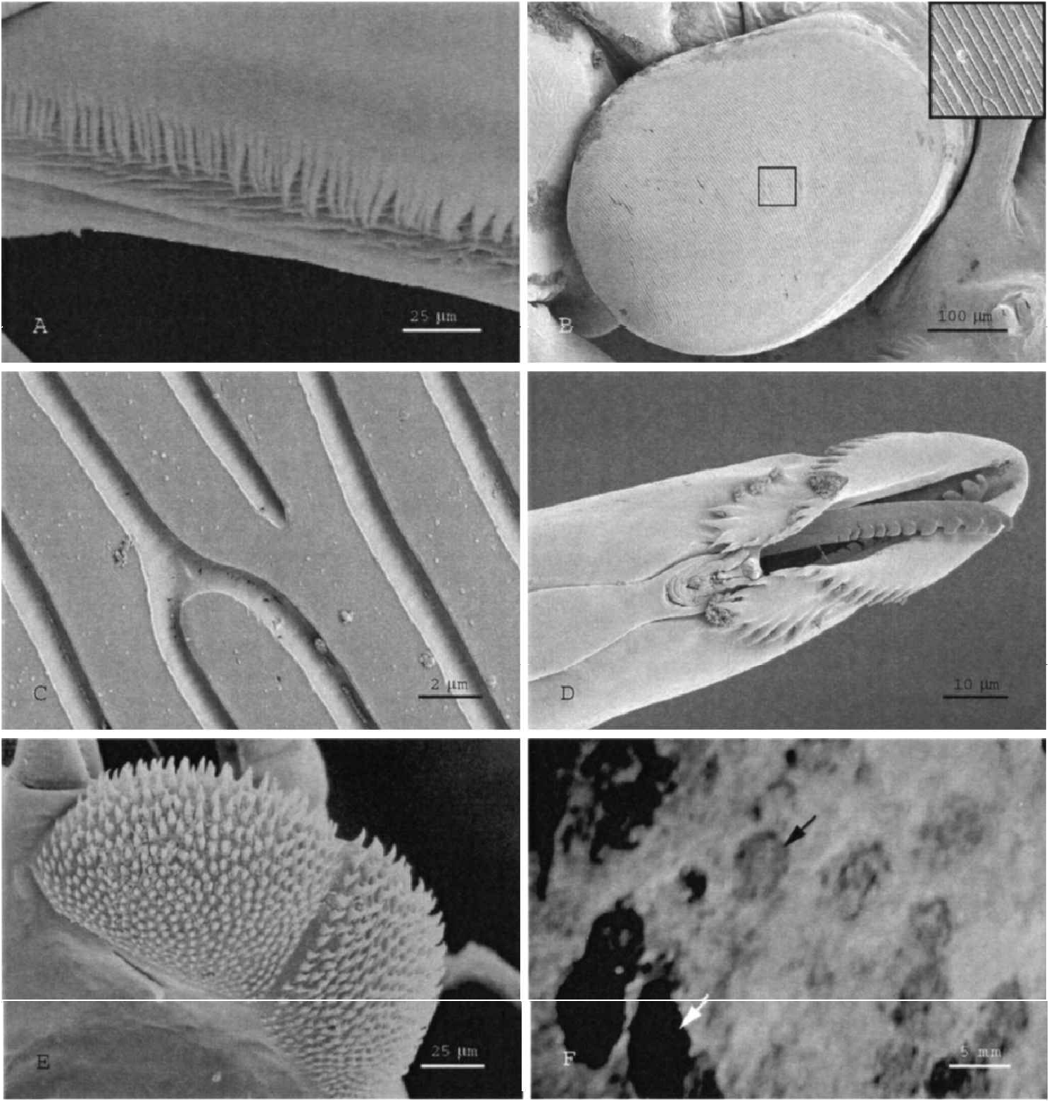
Body form shown in figure 1A View FIG and B View FIG . Length range 7.0-8.0 mm (mean = 7.6 mm, n = 10). Width range 3.83-4.28 mm (mean = 4.1 mm, n =10), with greatest width at cephalon, just anterior to the cephalon/thoracic junction. Height range 1.19-1.58 mm ( 1.4 mm, n =10). Although dorsal surface of body is smooth, pores are scattered across surface. Lateral margins of carapace are fl eshy and with frill (figure 2A).
Frontal plates well developed and narrow mesial extensions meet in midline. First pediger fused with head; hinder margin of cephalon with four or fi ve robust spines (sometimes heavily eroded), with another two on each extension of the cephalon. Dorsal thoracic plates on pedigers 2-4. Pediger 2: plates separate, extending beyond tip of pediger 3 almost to level of posterior limit of plates of pediger 4; straight posterior margin with four sharp spines. Plates of pediger 3 and 4 fused at their bases; posterior margins with shallow sinuses. Plates of pediger 4 extend over base of genital double somite. Genital double somite: almost circular; with well-de fi ned posterior projections (separated by a narrow sinus), each bearing an upturned triangular projection dorsally; often with a sub-marginal setospine either side near base of the sinus (but lacking in the holotype). Abdomen onesegmented covered dorsally by plate longer than wide and not extending to level of tips of caudal rami; margins at the greatest width of dorsal plate curved ventrally, giving the appearance, from dorsal aspect, of a slight projection: ventrally, joined broadly to genital double somite and posteriorly terminating in broad plate extending between bases of caudal rami. Caudal rami stout, curved, L-shaped in cross section; lateral surface is oblique proximally and follows line of abdomen to just beyond the widest point of dorsal abdominal plate beyond which level the caudal ramus is deflected outwards and tapers to a terminal spine which recurves slightly back towards midline of the animal. Upper margin of caudal ramus is sharply de fi ned beyond the stout spine, which marks the beginning of the curve outwards and carries a second smaller spine; ventral surface with tubercle near the proximo-lateral corner and with thin seta and small spine on the mesial edge.
Oral area. Adhesion pads present at bases of antennule, antennae and maxillipeds. The surface structure of a pad is illustrated in figure 2B View FIG and C View FIG . Pads also present anteriorly on lateral expansions of thoracomere 2. Antennule ( figure 1C View FIG ) of two articles: article 1 bearing 27 setospines, 21 stout and six small; article 2 bearing 12 naked, mostly curved, setae. Antenna of three articles ( figure 1D View FIG ): terminal article bearing large curved terminal spine and two spines marginally; article 2 with two ventral spines, one mid-article on broad base, the other at the distal margin. Mouth tube ( figure 1E View FIG ): of 10 females measured, oral cones 0.5-1.0 mm long, average 0.7 mm. Labrum ends in complex structure ( figure 2D View FIG ). Labium with two terminal fringes of backwardly directed denticles ( figure 2D View FIG ). Mandible ( figure 2D View FIG ), with slender shaft flattened and dentate near the tip. Maxillule ( figure 1F View FIG ) of two articles: basal article bearing 0 to two short setae; terminal article with large terminal, plus one small spine. Maxilla brachiform ( figure 1G View FIG ): article 1 (lacertus) unarmed; article 2 (brachium) without fl abellum, but with two distal spines, longer one fringed, shorter plumose; calamus bearing large claw with rows of spinules and apical patch of spinules. Maxilliped ( figure 1H View FIG ) of two articles: basal article (corpus maxillipedus) stout with nacreous-like pad; article 2 (subchela) unequally bilobed, with nacreous-like pad, which works against pad of article 1.
Legs View Table 1-4 biramose, each ramus of two articles, with spine and setal formula as follows:
Both rami of legs 1-3 ( figure 3A -C View FIG ) with two articles. Both rami of leg 4 (figure 3D) with one article; endopodite lacking spines. Leg 5 ( figure 3E View FIG ) consisting of outer seta and inner lobe with single terminal spine. Adhesion pads and denticulate areas illustrated in figure 3A -D View FIG . Figure 2E View FIG shows detail of a denticulate region.
Egg strings ( figure 3F View FIG ) slender, approximately same length as body, slightly curved. Eggs disc shaped.
Adult females vary in the extent of coloration. Colour patches dark chocolate - chestnut brown centrally shading outwards to transparent amber. Three colour patches occur on the cephalon, one anterior and triangular patches posterio-laterally. The considerable variation in the extent of separation to fusion between these three patches, resulting in considerable variation in the extent of amber-coloured areas about the eye spots, may be due to ontogenetic di ff erences. Colour patches also occur on frontal lobes; separately on segments 2 and 3 and at the bases of genital lobe projections; across most of segment 4 and abdominal segment (figure 1A).
Male ( figures 4, 5 View FIG View FIG )
Body form as in figure 4A View FIG and B View FIG . Length (not including setae on caudal rami) 5.2 -7.2 mm (mean = 6.0 mm, n =5), width 2.9-4.3 mm (mean = 3.3 mm, n = 5) and height 0.84-0.94 mm (mean = 0.90, n = 5). Cephalon rounded when viewed dorsally with head and fi rst pediger fused. Segment 2 bears two pairs of dorsal spines; segments 3, 4 and 5 each bear one pair of dorsal spines. Pedigers 2- 4 free, without dorsal plates except for lateral wing-like plates on pediger 2. Genital double somite with posterior corners terminating in prominent triangular projection. Coiled spermatophores visible within genital double somite. Abdomen two-segmented. Caudal ramus bearing four long, plumose setae and series of fi ne setules along inner margin. Oral area as in female except for distribution of adhesion pads. Adhesion pad ( figure 4B View FIG ) present at base of antennule ( figure 4C View FIG ) approximatel y half the length, and positioned at greater angle towards centre of the cephalon, than is the case in the female. Small adhesion pad situated on base of antenna ( figure 4D View FIG ). Absence of adhesion pads at base of maxilliped ( figure 4E View FIG ) and on lateral projections of pediger 2.
Legs View Table 1-4 biramose, each ramus of two articles, with spine and setal formula as follows:
Legs 1-4, figures 5A-D View FIG , which show the distribution of adhesion pads and denticulate areas. Leg 5 borne on genital double somite as lateral projection with three setae and one stout terminal spine. Leg 6 consisting of two setospines, outer longer than inner, borne on genital double somite near the origin of the abdomen.
Colour in life is pale pink and devoid of darker pigment.
Etymology
Rhincodonicus refers to the host of the copepods, the whale shark, Rhincodon typus .


| WAM |
Western Australian Museum |
No known copyright restrictions apply. See Agosti, D., Egloff, W., 2009. Taxonomic information exchange and copyright: the Plazi approach. BMC Research Notes 2009, 2:53 for further explanation.
|
Kingdom |
|
|
Phylum |
|
|
Class |
|
|
Order |
|
|
Family |
|
|
Genus |