
Hylica Stål
|
publication ID |
https://doi.org/10.11646/zootaxa.4388.4.4 |
|
publication LSID |
lsid:zoobank.org:pub:8042D16C-0564-4FEB-9921-EF37A3FF1A83 |
|
DOI |
https://doi.org/10.5281/zenodo.5980191 |
|
persistent identifier |
https://treatment.plazi.org/id/0381D703-FFE8-FFDE-FF52-FE6A471D4981 |
|
treatment provided by |
Plazi |
|
scientific name |
Hylica Stål |
| status |
|
Genus Hylica Stål View in CoL new record to China
Hylica Stål, 1863: 593 View in CoL ; Marschall, 1873: 367; Scudder, 1882: 156; Atkinson, 1885: 112; Distant, 1908: 252; Evans, 1946: 47; 1947: 165; Metcalf, 1962: 6; Oman et al., 1990: 218.
Type species: Hylica paradoxa Stål, 1863 , by original designation
Diagnosis. This genus can be easily distinguished from other genera of the subfamily Hylicinae by the strongly tuberculate head, pronotum and scutellum, and the oblong-obovate abdomen with dentate lateral margins.
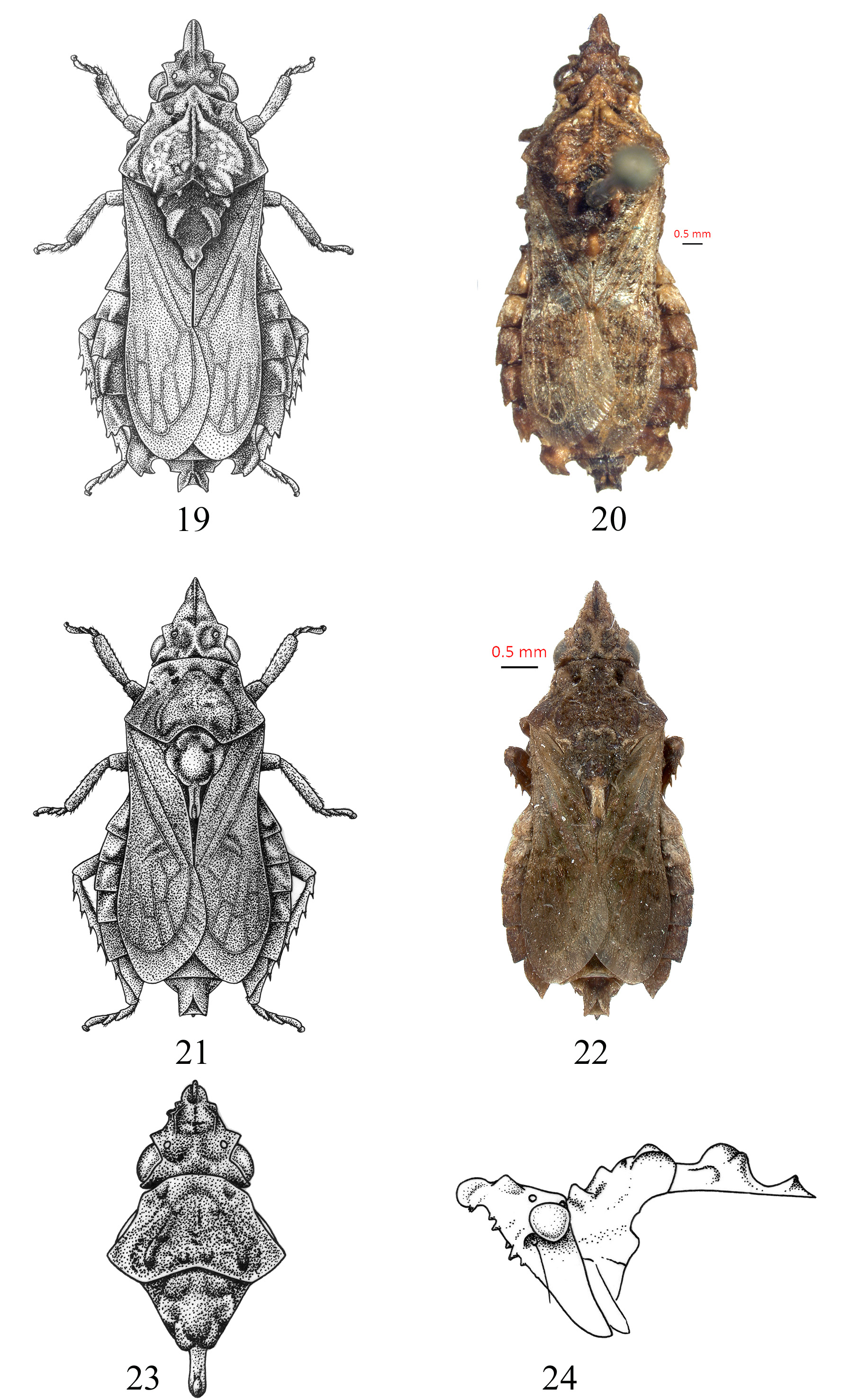
Description. Medium-sized to large leafhoppers (body length 6.6–11.5 mm incl. tegmen) with coloration brown-black. Body oblong-obovate and densely covered with many small dark and few light setae. Head, pronotum and scutellum strongly tuberculate. Vertex ( Figs. 4 View FIGURES 1–9 , 13 View FIGURES 10–18 , 23 View FIGURES 19–24. 19–20 ) acutely produced and resembling isosceles triangle slightly longer than wide with apical declivous process in lateral view; distal half of crown with median longitudinal ridge; ocelli situated on two tubercles of vertex near anterior angles of eyes, closer to eyes than to each other; pair of tubercles sinuated between ocelli and apex; tubercle in front of eye well developed and shelflike before eye in dorsal view; posterior margin straight and with pair of light spots. Face ( Figs. 6 View FIGURES 1–9 , 15 View FIGURES 10–18 ) irregular; frons bent forwards; frontoclypeal area slightly convex and with strongly developed muscle impressions; clypeal sulcus obvious; anteclypeus distinctly convex and extended slightly beyond gena; lorum small and narrow, well-separated from lateral margin of gena; gena with lateral margin evenly rounded below eye; almost completely concealing proepisternum; rostrum very short, just reaching base of front coxa; antenna shorter than length of crown; antennal ledge well developed. Pronotum ( Figs. 4, 5 View FIGURES 1–9 , 13, 14 View FIGURES 10–18 , 23, 24 View FIGURES 19–24. 19–20 ) backwardly elevated and sloping laterally, tuberculate; posterior margin with “w” shaped emargination. Exposed part of mesonotum and scutellum ( Figs. 7, 8 View FIGURES 1–9 , 16, 17 View FIGURES 10–18 , 23, 24 View FIGURES 19–24. 19–20 ) divided into three parts; anterior region disklike with some tubercles; middle region elevated with center sunken; posterior region lower than middle region, furnished near apex with conical tubercle and not reaching apex of clavus. Tegmina subcoriaceous; with four apical cells; appendix well developed and extended around wing apex. Abdomen ( Figs. 9 View FIGURES 1–9 , 18 View FIGURES 10–18 ) broader than tegmina at rest; strongly depressed and with lateral margins expanding into dentations. Legs somewhat short and usually with pale spots; front tibia above somewhat dilated and compressed; hind tibia above with spine-like setal bases; hind femur macrosetal formula 2+0+0; tibia with ca. 9, 8 setae in rows AD, PD, respectively.
Male genitalia: Pygofer ( Figs. 25–27, 32 View FIGURES 25–36. 25–31 , 37–39, 46, 47 View FIGURES 37–47. 37–42 ) lateral lobe without appendage, with many small stout setae, pygofer urn-shaped in dorsal view, base fused, terminal separated into two parts; genital valve almost degenerate and fused with subgenital plate; subgenital plate short, about half length of pygofer side, strawberryshaped; base of anal tube with pair of long, slender anal hooks almost reaching shaft apex of aedeagus. Base of style ( Figs. 31, 35 View FIGURES 25–36. 25–31 , 40, 45 View FIGURES 37–47. 37–42 ) elongate, apophysis of style very simple and short. Connective ( Figs. 31, 35 View FIGURES 25–36. 25–31 , 40, 45 View FIGURES 37–47. 37–42 ) Yshaped, stem longer than arms. Aedeagus ( Figs. 29, 30, 34, 36 View FIGURES 25–36. 25–31 , 41–44 View FIGURES 37–47. 37–42 ) with preatrium and dorsal apodeme, shaft almost straight, gonopore subapical on ventral surface.
Female genitalia. Female with sternite VII concave medially ( Figs. 48, 55 View FIGURES 48–61. 48–54 ). Ovipositor not protruding far beyond pygofer apex ( Figs. 49, 56 View FIGURES 48–61. 48–54 ). First valvula ( Figs. 50, 51, 57, 58 View FIGURES 48–61. 48–54 ) draped in middle and subapical region; width same throughout most of length, basal half short and nearly straight, distal half slightly curved upward; dorsal sculpturing pattern strigate to reticulate, submarginal for most of length. Second valvula ( Figs. 52, 53, 59, 60 View FIGURES 48–61. 48–54 ) curved; with toothed distal blade about or slightly more than half total length, apparently broadened, with or without a few small, somewhat irregular teeth. Third valvula ( Figs. 54, 61 View FIGURES 48–61. 48–54 ) with a row of small setae.
Ecology. This genus is arboreal and occurs generally in moist tropical forests although almost no observations have been made of live individuals.
Distribution. China, Nepal ( Bengal, Kathmandu), Borneo, Burma, India ( Assam), Thailand, Viet Nam, West Indonesia; Java, Laos ( Vientiane).


No known copyright restrictions apply. See Agosti, D., Egloff, W., 2009. Taxonomic information exchange and copyright: the Plazi approach. BMC Research Notes 2009, 2:53 for further explanation.
|
Kingdom |
|
|
Phylum |
|
|
Class |
|
|
Order |
|
|
Family |
|
|
SubFamily |
Hylicinae |
Hylica Stål
| Tang, Jiu & Zhang, Yalin 2018 |
Hylica Stål, 1863 : 593
| Stål, 1863 : 593 |
| Marschall, 1873 : 367 |
| Scudder, 1882 : 156 |
| Atkinson, 1885 : 112 |
| Distant, 1908 : 252 |
| Evans, 1946 : 47 |
| Metcalf, 1962 : 6 |
| Oman et al ., 1990 : 218 |