
Rhyncaphytoptus miliusius, Lv & He & Gao & Tan & Wang, 2022
|
publication ID |
https://doi.org/10.11646/zootaxa.5175.5.2 |
|
publication LSID |
lsid:zoobank.org:pub:9D012CAB-B3A3-4001-9DFE-AA44780A7E4C |
|
DOI |
https://doi.org/10.5281/zenodo.7009438 |
|
persistent identifier |
https://treatment.plazi.org/id/0385565F-9326-FFAB-FF33-F89A4B6AFADD |
|
treatment provided by |
Plazi |
|
scientific name |
Rhyncaphytoptus miliusius |
| status |
sp. nov. |
Rhyncaphytoptus miliusius sp. nov.
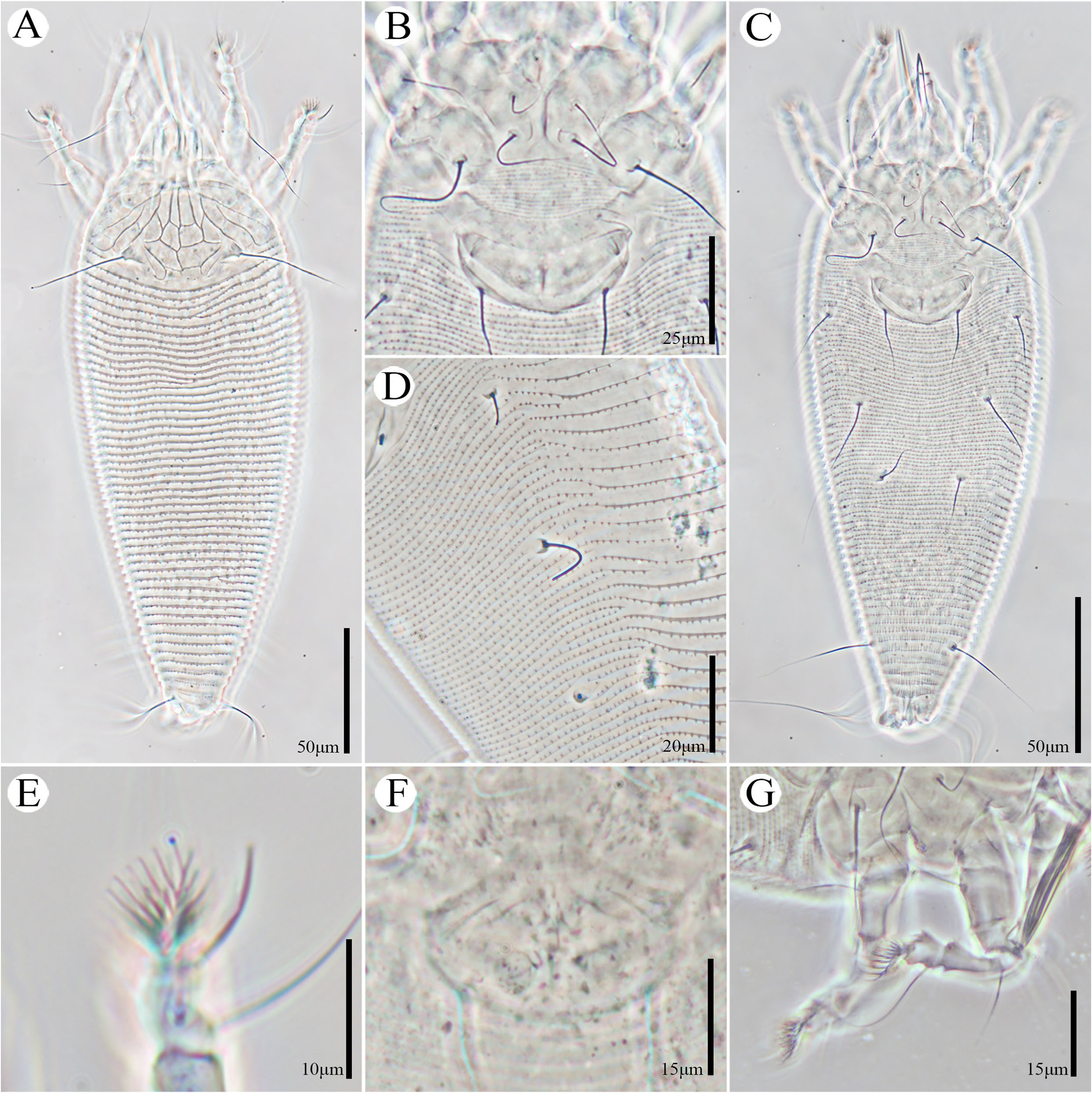
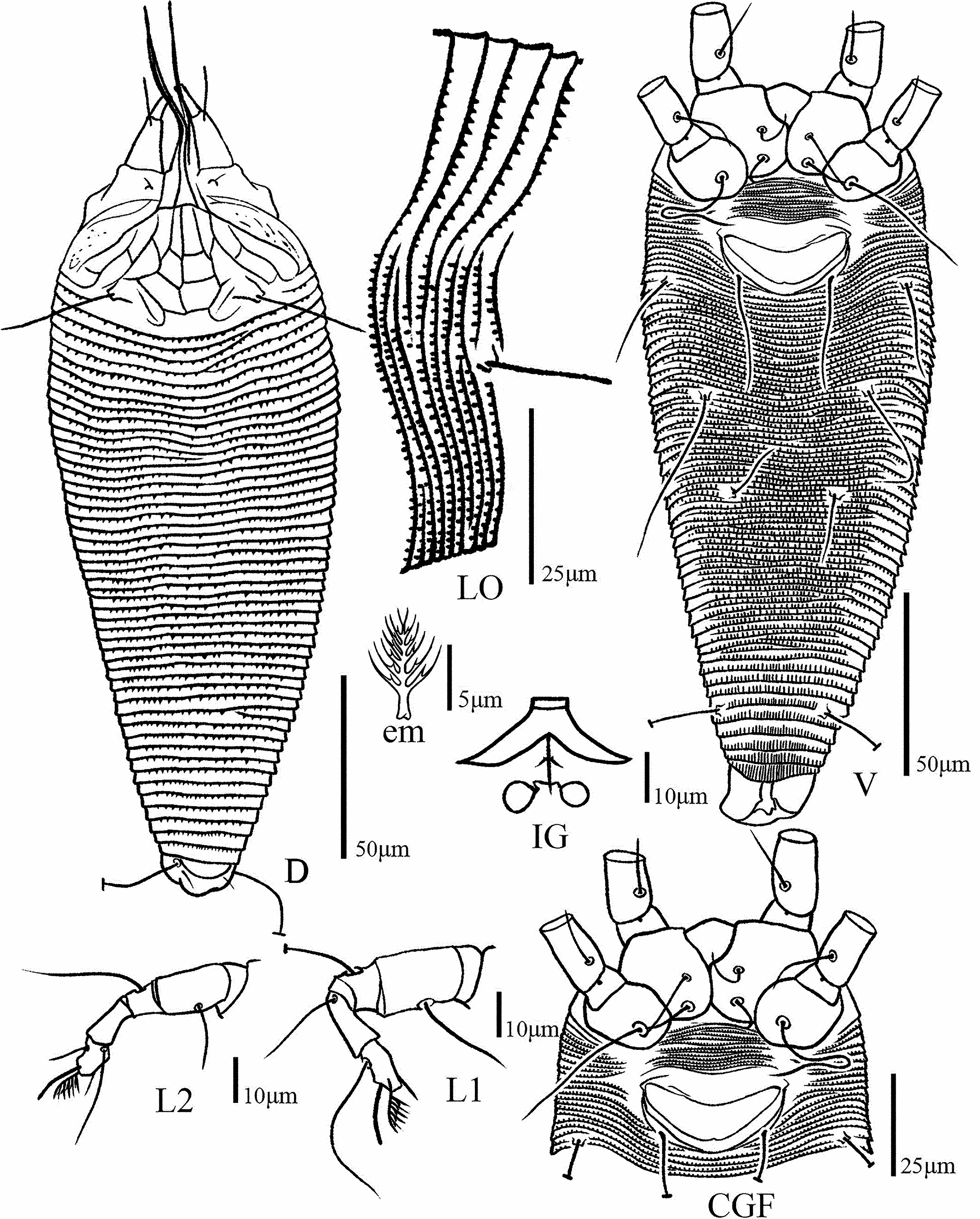
( Figs 4–5 View FIGURE 4 View FIGURE 5 )
Diagnosis. Body fusiform, yellow. Gnathosoma large in comparison to body, empodium entire, prodorsal shield lobe present. Median line incomplete, absent at basal 1/5, admedian lines and submedian lines complete; median and admedian lines contected with two transverse lines forming six cells. Scapular tubercles on rear shield margin, scapular setae ( sc) directed backwards. Coxal area smooth, prosternal apodeme present. Legs with series of setae, tarsal empodium entire, 7-rayed. Female genital coverflap smooth. Dorsal annuli 50 (50–54), ventral annuli 94 (92–96).
Description. Female (n=10).
Body. Fusiform ( Figs. 4A, 4C View FIGURE 4 , 5D, 5V View FIGURE 5 ), yellow in color. 214 (208–221) long, 76 (74–78) wide, 71 (69–74) thick. Gnathosoma. Projecting downwards 46 (42–48), pedipalp coxal setae ( ep) 5 (4–5), dorsal pedipalp genual setae ( d) 11 (8–12), unbranched, subapical pedipalp tarsal setae ( v) 3*, cheliceral stylets 55 (52–58). Prodorsal shield. 30 (29–32) long, 58 (56–60) wide, frontal lobe present. Median line incomplete, median line absent at basal 1/5, admedian lines complete, connected to median line by three cross lines at rear ¼, ½ and ¾ producing six small cells, base forming arc. Submedian lines complete. Scapular tubercles set ahead of rear shield margin, 33 (31–34) apart, scapular setae ( sc) 31 (28–34), scapular setae directed posterolaterally. Coxisternal plates ( Figs. 4B View FIGURE 4 , 5 View FIGURE 5 CGF). Prosternal apodeme 9 (9–12), coxal area smooth, anterolateral setae on coxisternum І ( 1b) 11 (10–13), 12 (10–13) apart; proximal setae on coxisternum І ( 1a) 22 (20–24), 11 (10–13) apart; proximal setae on coxisternum ІІ ( 2a) 36 (35–39), 34 (32–37) apart. Coxigenital annuli 15 (14–15).
Legs ( Figs. 4G View FIGURE 4 , 5L View FIGURE 5 1 View FIGURE 1 , 5L View FIGURE 5 2 View FIGURE 2 ). Segments normal. Legs I 41 (40–46), trochanter4 (3–5), femur 13 (12–14),basiventral femoral setae ( bv) 16 (15–18); genu 6 (5–7), antaxial genual setae ( l′ ′) 31 (30–33); tibia 11 (9–13), paraxial tibial setae ( l′) 13 (12–14), setae located ¼ from dorsal base; tarsus 6 (6–8), paraxial fastigial tarsal setae ( ft′) 24 (21–25), antaxial fastigial tarsal setae ( ft′ ′) 28 (26–30), paraxial unguinal tarsal setae ( u′) 5 (4–5); tarsal empodium ( em) 8 (7–8), simple, 7-rayed, tarsal solenidion ( ω) 9 (9–10). Legs ІІ 34 (31–36), trochanter 4 (3–4), femur 11 (11–13), setae ( bv) 15 (13–17); genu 6 (5–6), setae ( l′ ′) 14 (14–16); tibia 9 (9–12); tarsus 7 (6–9), setae ( ft′) 8 (7–11), setae ( ft′ ′) 26 (25–29), setae ( u′) 5 (4–5); tarsal empodium ( em) 8 (7–8), simple, 7-rayed, tarsal solenidion ( ω) 9 (8–10).
Opisthosoma ( Figs. 4A, 4B View FIGURE 4 , 5D, 5V View FIGURE 5 ). Dorsally arched, dorsal annuli 50 (50–54), with sharp-angled or spine microtubercles. Ventral annuli 94 (92–96), with round microtubercles; Setae c2 23 (22–24), 64 (63–64) apart, on ventral annulus 24 (23–24); setae d 52 (49–55), 43 (43–44) apart, on ventral annulus 40 (40–41); setae e 20 (18–24), 26 (25–27) apart, on ventral annulus 58 (56–60); setae f 41 (39–45), 28 (27–30) apart, on 6 (6–7) ventral annulus from rear; setae h1 5 (5–7), 10 (9–11) apart; setae h2 65 (60–69), 15 (15–16) apart. Female genitalia ( Figs. 4B View FIGURE 4 , 5 View FIGURE 5 IG). 15 (14–17), 34 (33–36) wide, coverflap smooth and bowl shaped, setae 3a 35 (32–37), 24 (23–24) apart. Internal genitalia: anterior transverse genital apodeme trapezoidal in ventral view, distally folded, spermathecal tubes relatively short, oblique apodeme present under the anterior genital apodeme, spermatheca almost globose or ovoid, directed latero-posterad.
Male unknown.
Type material. Holotype female (slide number Jinzhongshan 2022.2~2.1, GXU), Jinzhongshan Nature Reserve , Baise City, Guangxi Zhuang Autonomous Region, China ( 24°36′07′′N, 104°52′37′′E, 1000 m), 3 May 2021, from Miliusa sinensis Finet & Gagnep. (Annonaceae) . Coll. Mengchao Tan, Liangxin Liu and Ankang Lv. GoogleMaps Paratypes nine females (slide number Jinzhongshan 2022.2.2 ~2.10, GXU), same data as holotype.
Host plant. Miliusa sinensis Finet & Gagnep. (Annonaceae) .
Relation to the host plant. The mites are vagrants on the undersurface of the leaves, no visible damage was observed.
Distribution. China ( Guangxi).
Etymology. The specific designation miliusius is derived from the generic name of the type host plant, Miliusa ; feminine in gender.
Remarks. The new species is similar to Rhyncaphytoptus castanifoliae Keifer, 1940 found on Castanea dentata (Marshall) Borkh. Chestnut , ( Fagaceae ) which is characterized as follows: median line incomplete, coverflap smooth, opisthosoma setae h1 present. But can be differentiated from the latter by having the ventral annuli with sharp-angled microtubercles ( vs. dorsal annuli with dash-like microtubercles in R. castanifoliae ), tarsal empodium ( em) 7-rayed ( vs. tarsal empodium ( em) 5-rayed in R. castanifoliae ), the length of scapular setae sc 31 μm ( vs. 12 μm in R. castanifoliae ). The new species is also similar to Rhyncaphytoptus acer Chen, Wei & Qin, 2004 found on Acer davidii Franch. (Aceraceae) which is characterized as follows: admedian lines complete, coverflap smooth, prosternal apodeme present. But can be differentiated from it by tarsal empodium ( em) 7-rayed ( vs. tarsal empodium ( em) 6-rayed in R. acer ), the length of scapular setae sc 31 μm ( vs. 17.5 μm in R. acer ). The new species is also similar to Rhyncaphytoptus spinus Li, Xue & Hong, 2012 found on Lonicera rupicola Hook. F. et Thoms. (Caprifoliaceae) which is characterized as follows: coverflap smooth, prosternal apodeme and opisthosoma setae h1 present. But can be differentiated it by dorsal annuli with sharp-angled microtubercles ( vs. dorsal annuli with long spiny microtubercles in R. spinus ), the length of scapular setae sc 31 μm ( vs. 21 μm in R. spinus ), the length of opisthosomal setae d 52 μm ( vs. 97 μm in R. spinus ). The main differences lie in the following Table 2 View TABLE 2 .


No known copyright restrictions apply. See Agosti, D., Egloff, W., 2009. Taxonomic information exchange and copyright: the Plazi approach. BMC Research Notes 2009, 2:53 for further explanation.
|
Kingdom |
|
|
Phylum |
|
|
Class |
|
|
Order |
|
|
Family |
|
|
SubFamily |
Rhyncaphytoptinae |
|
Genus |