
Agnosthaetus affinis Clarke
|
publication ID |
https://doi.org/10.1649/0010-065X-65.mo4.1 |
|
publication LSID |
lsid:zoobank.org:pub:0818A3A2-AB42-43D8-8F76-4F65F367C584 |
|
persistent identifier |
https://treatment.plazi.org/id/DA354009-37D1-46A7-9792-616E8E97323F |
|
taxon LSID |
lsid:zoobank.org:act:DA354009-37D1-46A7-9792-616E8E97323F |
|
treatment provided by |
Carolina |
|
scientific name |
Agnosthaetus affinis Clarke |
| status |
sp. nov. |
(34) Agnosthaetus affinis Clarke View in CoL , new species
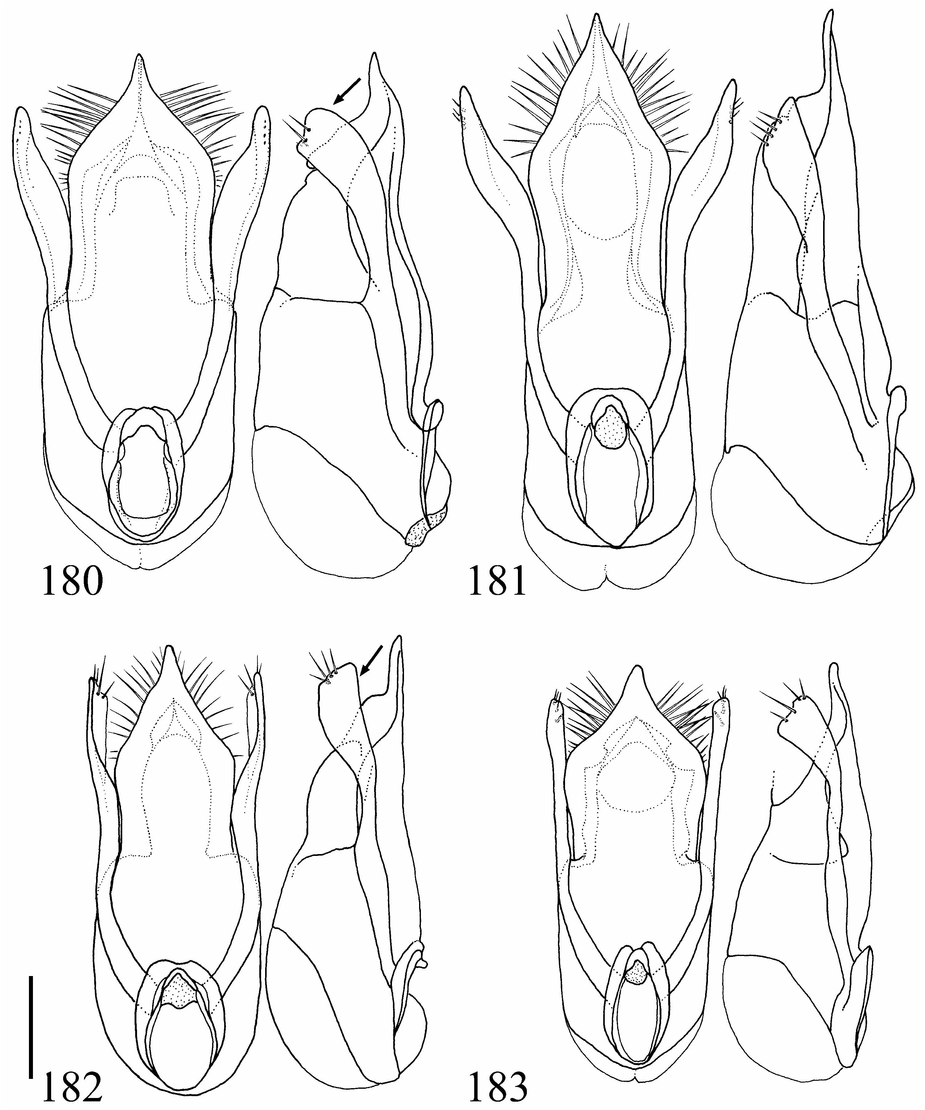
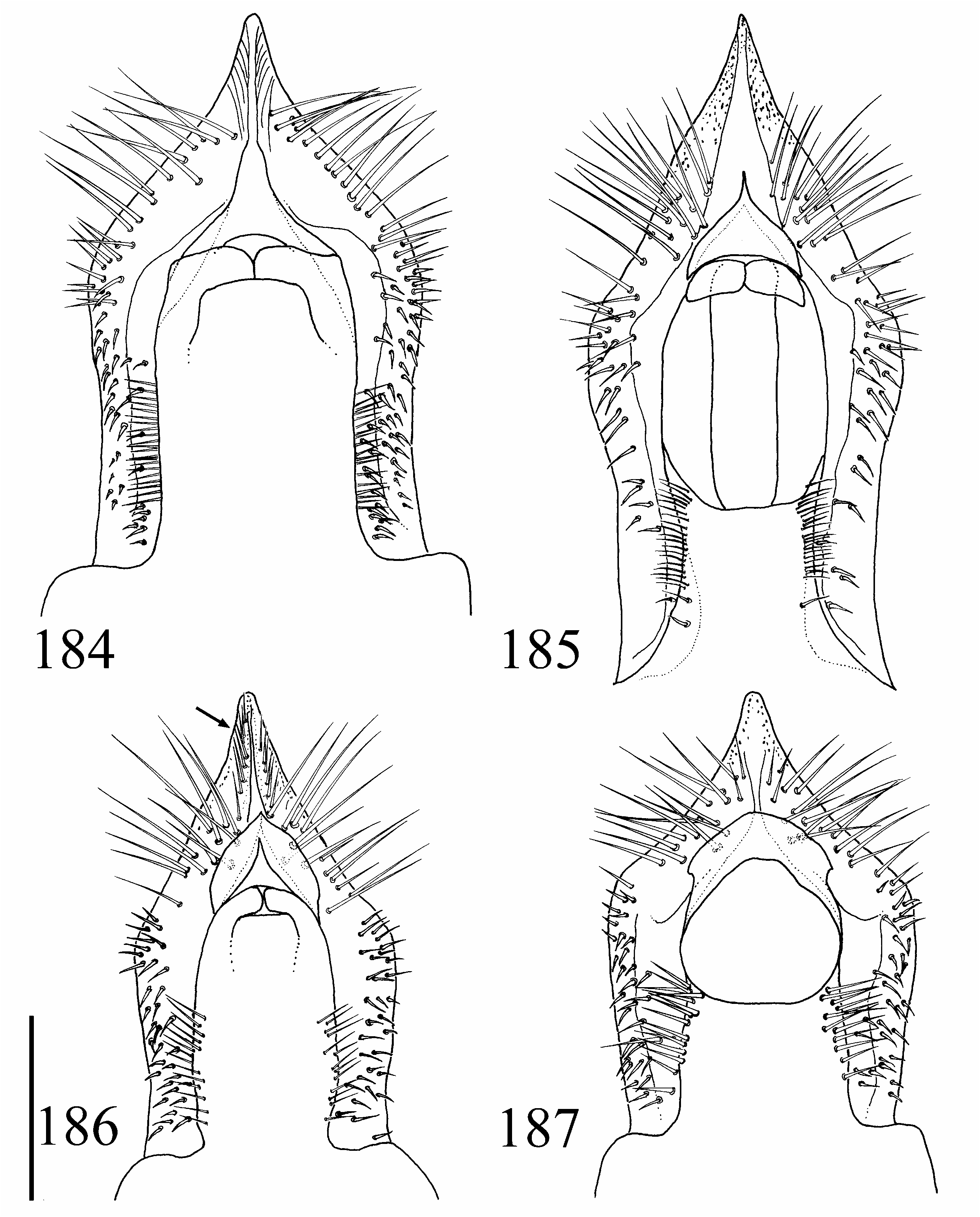
( Figs. 179 View Figs , 183 View Figs , 187 View Figs , Map 7 View Map 7 )
Type Material. Holotype. ♂, with three labels: “ NEW ZEALAND: CO| Piano Flat [ 45°32′S, 169°1′E], Titan Rocks| track 9-XII-1998 | C. Hall ex. litter/ SM0673434 | KUNHM-ENT / HOLOTYPE Agnosthaetus affinis Clarke , ♂, design. D. Clarke 2011”, in KSEM GoogleMaps . Paratypes. 6 specimens ( 5♂ 1♀). Same data as holotype GoogleMaps : 5♂, KUNHM-ENT SM0673340, KUNHM-ENT SM0673398, KUNHM- ENT SM0673414, KUNHM-ENT SM0673428, KUNHM-ENT SM0673430, 1♀, KUNHM-ENT SM0673427 (in KSEM) .
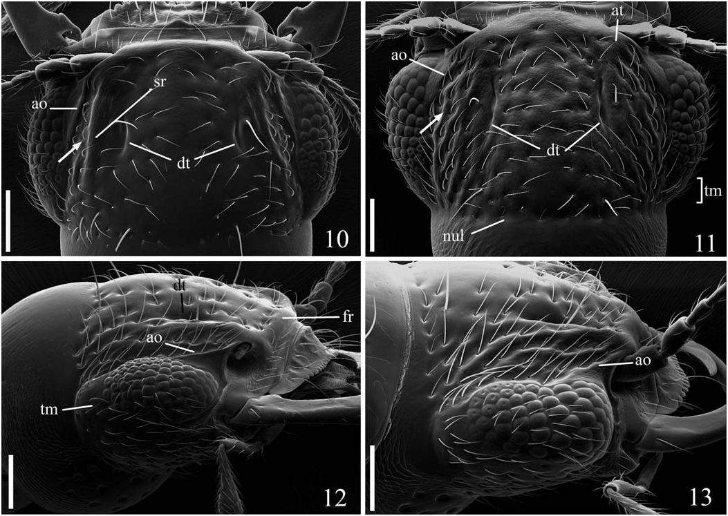
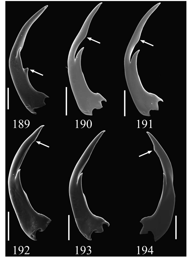
Diagnosis. The distinctive and even microsculpture on the head and thorax of A. affinis distinguishes this species from others in the nunni species-group, but makes it closely resemble species in the thayerae and truncatus speciesgroups, as well as A. lanceolatus (North Island) and A. ecarinatus . From these species, A. affinis may be distinguished by the combination of distinct antenno-ocular carina ( Fig. 10 View Figs , ao; which alone distinguishes it from A. ecarinatus ), more or less distinct sublongitudinal ridge ( Fig. 10 View Figs , sr; which distinguishes it from the truncatus speciesgroup), shallow and sparse dorsal head punctuation, elytra with single lateral ridge ( Fig. 24 View Figs , ek) (these last two easily distinguish it from the thayerae species-group), and males with unmodified labral teeth ( Fig. 179 View Figs ; distinguishes males from all aforementioned species except A. ecarinatus ).
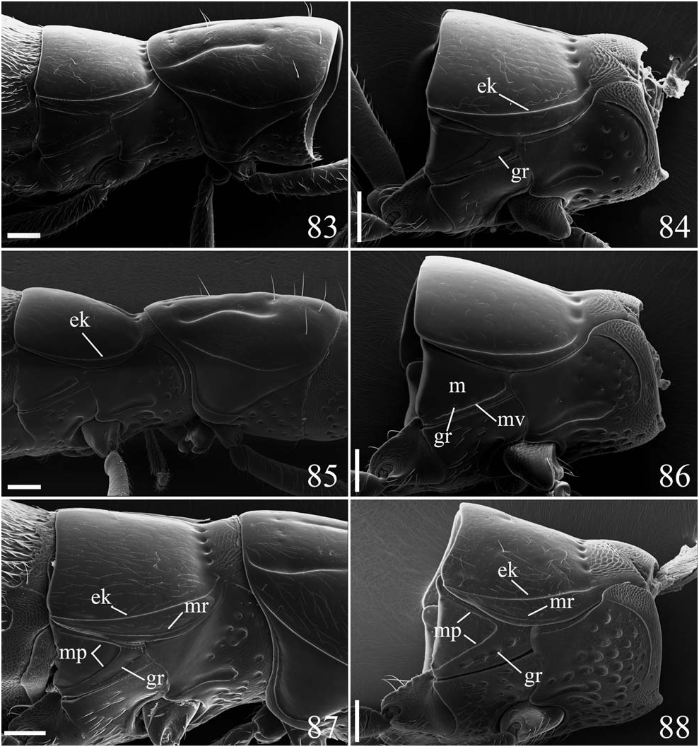
Description. Color: More or less uniformly yellowish brown, with abdominal segment VI distinctly darker, nearly black ( Fig. 5 View Figs ). Head: Frontal ridge absent. Dorsum sparsely punctate; with punctures distributed anteriorly, laterally, and posteriorly on disc, middle part impunctate. Punctures shallow, rather indistinct; diameter greater than diameter of eye facet; interpuncture distance approximately 0.5–1.0X puncture diameter. Dorsal microsculpture present on entire or most of surface; distinctly reticulate, fainter posteriorly. Dorsal tentorial sulcus ( cf. Figs. 10–11 View Figs , dt) narrowly ovate; width subequal to or slightly greater than puncture diameter. Sublongitudinal ridge ( cf. Fig. 10 View Figs , sr) distinct; not confused by smaller carinae or punctures ( cf. Fig. 10 View Figs , sr); crest at antennal tubercle with distinct microsculpture. Area above and behind antenno-ocular carina ( Figs. 10–11 View Figs , arrow) more or less smooth, without subsidiary carinae. Antenno-ocular carina joining eye at or behind middle ( cf. Fig. 10 View Figs , ao). Temple ( Fig. 11 View Figs , tm) short, less than 50% EYL. Subocular surface more or less evenly microsculptured ( cf. Fig. 65 View Figs ). Labrum not distinctly sexually dimorphic ( Fig. 179 View Figs ). Apical labral margin in males very shallowly emarginate medially, evenly dentate, with 16–21 teeth ( n =4), all teeth normal, projecting more or less anteriorly. Apical labral margin in females broadly convex, not emarginate medially; with 17 teeth ( n =1), all teeth subequal in length. Adoral labral surface in males smooth, without subapical transverse ridge. Mandible sexually dimorphic; males with single, dorsally directed tooth, without preapical spur ( cf. Fig. 189 View Figs ); females with single, mesially projecting tooth, without spur. Prothorax: Pronotum with distinctly reticulate microsculpture. Medial pronotal sulci anteriorly continuous with anterior punctures ( cf. Fig. 76 View Figs ). Distance between medial sulci very slightly greater posteriorly. Pronotal basolateral carina distinct ( cf. Fig. 76 View Figs , bp). Pronotal macrosetal punctures distinct ( cf. Fig. 73 View Figs ). Medial pronotal seta subequidistant from medial and lateral sulci ( cf. Fig. 73 View Figs , mu). Pronotal hypomeron ( Fig. 24 View Figs , hy) with distinct reticulate microsculpture. Prosternum with distinctly reticulate microsculpture. Pterothorax: Elytron ( Fig. 23 View Figs , e) with distinct microsculpture; with 2 macrosetae or with 3 macrosetae, set in distinct punctures; laterally with single ridge ( cf. Fig. 84 View Figs , ek). Mesothoracic epimeral region ( Fig. 24 View Figs , mer) with distinct microsculpture. Metathoracic pleural region ( Fig. 24 View Figs , m) with distinct reticulate microsculpture. Metathoracic pleural ridge fully developed ( cf. Fig. 24 View Figs , mp); metathoracic pleural groove ( Fig. 24 View Figs , gr) incomplete posteriorly, forming elongate oval punctiform impression. Abdomen: Abdominal vestiture short, somewhat appressed, dorsally more or less evenly projecting posteriorly but with middle setae directed posteromedially. Abdominal sternite V of male with surface impressed apico- medially, flanked by coarse acuminate setae forming small tufts apically, apex of sternite not distinctly sinuous; VI with subapicomedial patch of denser setae. Aedeagus ( Fig. 183 View Figs ): “ Type B” (see description on p. 8). Apical part of median lobe slightly narrower basally, not forming distinct lateral lobes; produced concavely into narrow point. Apicolateral setae small; apicomedial setae up to 10X longer than apicolateral setae ( Fig. 187 View Figs ). Paramere not exceeding apex of median lobe; in lateral view produced apically into lobe; with apical part perpendicular to median lobe; in dorsal view with outer side more or less straight; with 2 small setae at apex and 2 large ones on ventral edge.
Etymology. The specific epithet affinis is an adjective from the Latin affinis (-e), near or related to, for its general similarity to other species in both the nunni species-group, and those other species with distinctive microsculpture.
Distribution. ( Map 7 View Map 7 ). South Island: CO.


No known copyright restrictions apply. See Agosti, D., Egloff, W., 2009. Taxonomic information exchange and copyright: the Plazi approach. BMC Research Notes 2009, 2:53 for further explanation.
|
Kingdom |
|
|
Phylum |
|
|
Class |
|
|
Order |
|
|
Family |
|
|
Genus |