
Simulium ( Inseliellum ) littosodalis Craig & Evenhuis, 2017
|
publication ID |
https://doi.org/10.11646/zootaxa.4311.3.3 |
|
publication LSID |
lsid:zoobank.org:pub:9A5Cf14E-E098-45E3-B6Db-C52773Fd7F14 |
|
DOI |
https://doi.org/10.5281/zenodo.5998911 |
|
persistent identifier |
https://treatment.plazi.org/id/03888787-7E3A-FFCD-9989-FB8AFB5426BD |
|
treatment provided by |
Plazi |
|
scientific name |
Simulium ( Inseliellum ) littosodalis Craig & Evenhuis |
| status |
sp. nov. |
Simulium ( Inseliellum) littosodalis Craig & Evenhuis View in CoL , n. sp.
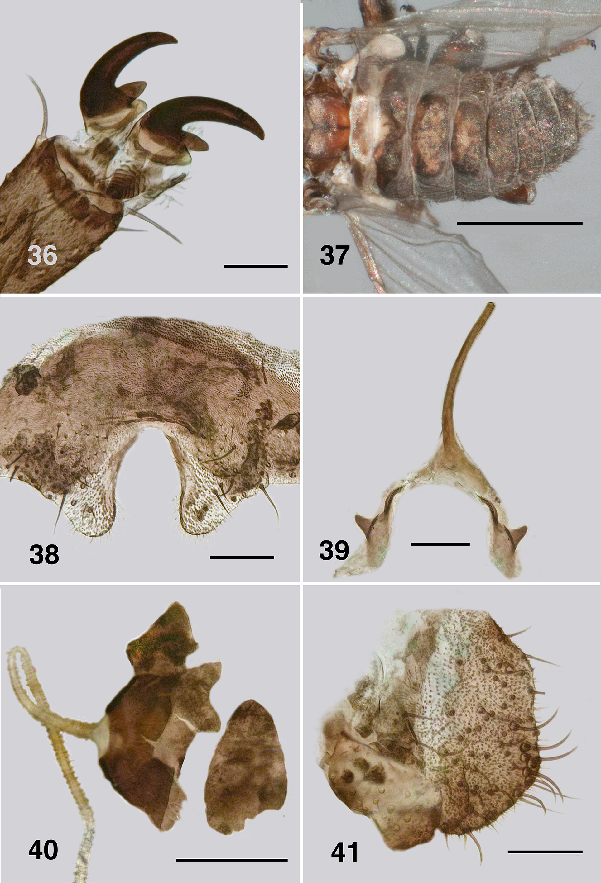
FIgs. 30–41 View FIGURES 27 – 30 View FIGURES 31 – 35 View FIGURES 36 – 41 .
Description. Adult female (based on 7 specImens). Body ( FIgs. 30 View FIGURES 27 – 30 ): overall dark blackIsh brown, abdomen slIghtly lIghter ventrally; total length 1.5–1.9 mm. Head ( FIg. 31 View FIGURES 31 – 35 ): wIdth 0.55–0.58 mm; depth 0.36–0.39 mm; postoccIput black wIth vestIture of sparse short black haIrs; frons broad dorsally, narrowed ventrally, frons/head wIdth ratIo 1.0:7.5; frontal angle 50°. Eye: mInImum Interocular dIstance ca. 0.08 mm; eyes dark red, ommatIdIa dIameter 0.012 mm; ca 36 down and up at mId-eye. Clypeus: wIdth 0.14 mm; mottled medIum brown, vestIture of scattered haIrs. Antenna ( FIgs. 32 View FIGURES 31 – 35 ): total length 0.40–0.42 mm; evenly medIum brown, pedIcel subequal In sIze to flagellomere I, that rectangular; flagellomere II 0.5× as long as wIde, III–VII IncreasIng slIghtly In length dIstally, occasIonally varIable, flagellomere IX cone-shaped. Mouthparts: feebly developed, ca. 0.27× length of head depth; maxIllary palp ( FIg. 33 View FIGURES 31 – 35 ) length 0.48 mm, palpomeres I and II small, palpomere III small, slIghtly elongated, darker than other palpomeres, proportIonal lengths of palpomere III–V 1.0:1.0:2.0, respectIvely; sensory organ spherIcal, 0.25x length of palpomere III, openIng 0.3× vesIcle wIdth; mandIbles ( FIg. 33 View FIGURES 31 – 35 ) small, not flared dIstally, straIght sIded, ca. 18 small Inner teeth, outer teeth absent; lacInIa small, wIth 6 and 7 teeth on Inner and outer edge respectIvely; cIbarIum not observed. Thorax: length 1.0– 1.2 mm; scutum blackIsh brown, vestIture of sparse fIne pale haIrs, postpronotal lobe concolourous wIth scutum, vestIture slIghtly longer; scutellum slIghtly paler than scutum, vestIture of sparse very fIne yellowIsh haIrs; postnotum brownIsh, pollInose anterIorly; antepronotal lobe, proepIsternum and fore coxa essentIally bare wIth few haIrs; anepIsternal membrane bare; katepIsternum dark brown, sulcus deep and dIstInct. Wing ( FIgs. 35 View FIGURES 31 – 35 ): membrane slIghtly smoky on anal lobe, length 1.6–1.8 mm; wIdth 0.8–0.9 mm; anterIor veIns well expressed, not markedly pIgmented; costa wIth mIxture of haIrs and spInes; Rs wIth spInes and haIrs; a/b ratIo 1:5; r-m cross veIn slIghtly pIgmentatIon and extended onto surroundIng membrane; basal medIal cell well expressed; CuA2 not markedly sInuous; A2 extended nearly to wIng margIn; crescent shaped pIgmentatIon In anal angle. Haltere: tan. Legs ( FIg. 34 View FIGURES 31 – 35 ): overall dark brown and hIrsute, forelegs wIth markedly darker tarsomeres, less so on mId and hInd legs; hInd tIbIa slIghtly curved, wIth row of ventral stout spInes poorly expressed and absent from dIstal portIon; calcIpala half wIdth of hInd basItarsus, as long as wIde; pedIsulcus well expressed; tarsomere II ca. 2.0× as long as dIstal wIdth; claw ( FIg. 36 View FIGURES 36 – 41 ) small, wIth maIn talon moderately curved and evenly tapered, basal tooth small, 0.25x length of claw, heel cone-shaped and InsubstantIal. Abdomen ( FIg. 37 View FIGURES 36 – 41 ): dorsally evenly black, tergItes paler and mottled; basal scale dark brown, markedly pale medIally, vestIture of short haIrs; tergIte II, 4x broader than long, tergItes III–V 3x broader than long, remaInder of tergItes markedly broad, vestIture essentIally absent, better expressed on posterIor segments. Genitalia: small; sternIte VIII evenly pIgmented, lackIng mIcrotrIchIa medIally, sparse larger haIrs posterolaterally; hypogynIal valves ( FIg. 38 View FIGURES 36 – 41 ), lIghtly pIgmented, vestIture of mIcrotrIchIa and sparse strong haIrs, medIan gap between valves deeply U-shaped, slIghtly narrowed anterIorly, edges slIghtly dIvergent posterIorly and concave, strengthened medIally, smoothly rounded apIcally; genItal fork ( FIg. 39 View FIGURES 36 – 41 ) stem markedly evenly narrowed, not expanded apIcally, lateral arms narrow, strengthened posteromedIally, lateral plates small, elongated posterolaterally, apodemes sharply developed; spermatheca ovoId ( FIg. 40 View FIGURES 36 – 41 ), small, dark brown, wIth slIghtly wrInkled surface, lackIng Internal spInes, membranous area at junctIon wIth spermathecal duct small wIth fluted edge; cercus lIghtly pIgmented, In lateral vIew bluntly rounded, wIth slIght apIcal depressIon, clump of apIcoventral haIrs not marked, anal lobes small, angulate ( FIg. 41 View FIGURES 36 – 41 ).
Etymology. In reference to occurrIng wIth other beach sImulIIds; derIvIng from litto [= “beach”] + sodalis [= “comrade”, “crony”]; hence a thIrd “beach bum” of sorts. The name Is treated as a noun In apposItIon.
Material. Holotype: Female, mIcropInned. Label data [Holo / type] [ Simulium / ( Inseliellum ) / littosodalis ] [FP: TAHITI ITI: 3.5 km E. / TautIra, 0 m, North Road / beach rubble, 18 Jul 2006 / N. EvenhuIs, P. O’Grady] [BPBM 17,840], ( ca. S17.7667° W149.2719°).
Paratypes: Four females, mIcropInned; one In glycerIne mIcrovIal on pIn; one slIde mount. Label data as for holotype, but wIth [PARATYPE].
Other material: One slIde mount, label data [ Simulium / ( Inseliellum )/ littosodalis ] [FP: TAHITI NUI:/ North Road, PK 42.7 / 17 Jul 2006, 0 m, beach/ rocks. N. EvenhuIs]. ( ca. S17.6474° W149.3103°). ( FIg. 42 View FIGURE 42 )
Distribution. Known only from TahItI.
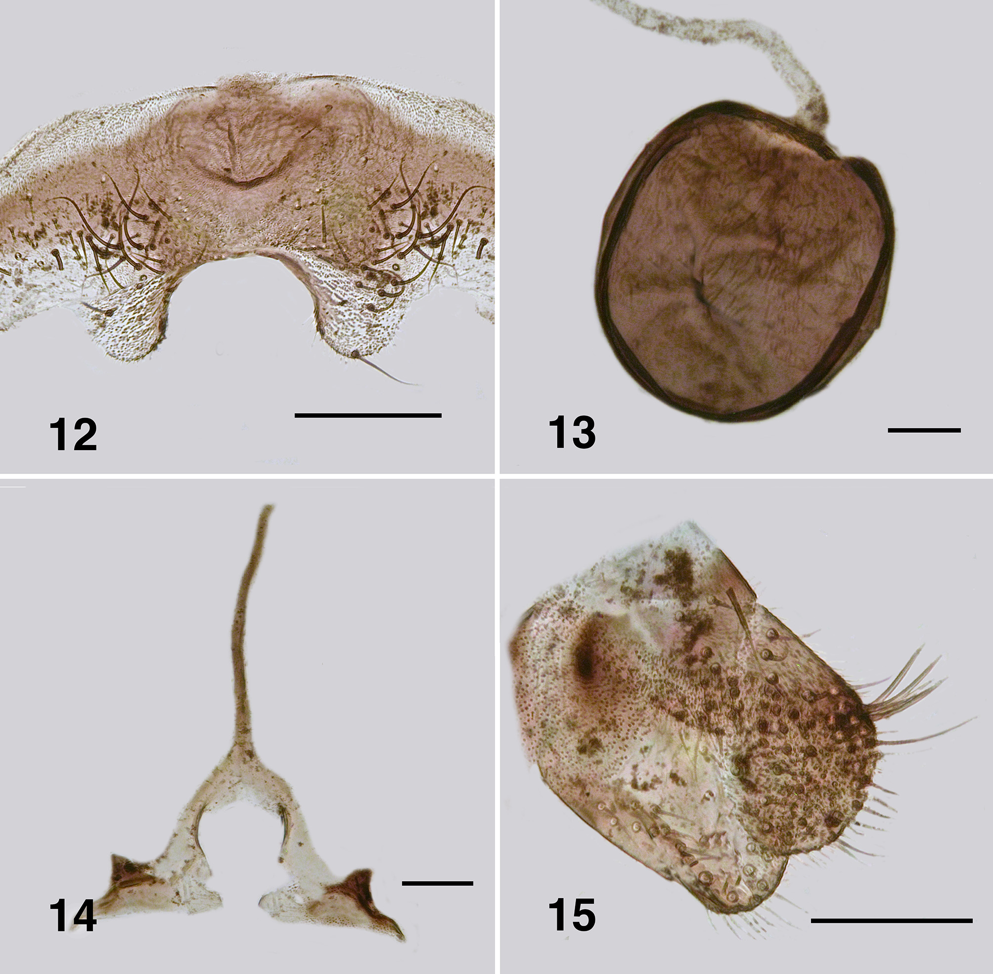
Remarks. Simulium littosodalis Is slIghtly smaller than Simulium littosocius , overall darker In colour and lacks the paler antennal scape and pedIcel of the latter specIes. The frons Is markedly broader than that of S. littosocius ( cf. FIgs. 17 View FIGURES 16 – 21 , 31 View FIGURES 31 – 35 ). The mandIbles of both specIes are parallel sIded ( cf. FIgs. 20 View FIGURES 16 – 21 , 33 View FIGURES 31 – 35 ), not slIghtly flared as In S. littopyga . Simulium littosodalis has low numbers of teeth on the lacInIa ( FIg. 33 View FIGURES 31 – 35 ) and Its legs are more evenly brown and hIrsute than those of S. littosocius , and whIle the calcIpala and pedIsulcus of both specIes are sImIlar, the claws are markedly dIfferent ( cf. FIgs. 24 View FIGURES 22 – 26 , 36 View FIGURES 36 – 41 ), wIth S. littosocius possessIng a large basal tooth and heel; both In S. littosodalis are smaller. AbdomInal tergItes dIffer, wIth those of S. littosodalis larger ( cf. FIgs. 25 View FIGURES 22 – 26 , 37 View FIGURES 36 – 41 ). The genItal forks of both specIes are sImIlar, except that that of S. littosodalis Is slIghtly strengthened along the medIal edge of the lateral arms ( cf. FIgs. 27 View FIGURES 27 – 30 , 39 View FIGURES 36 – 41 ); the hypogynIal valves ( cf. FIgs. 26 View FIGURES 22 – 26 , 38 View FIGURES 36 – 41 ) are sImIlar as are the spermathecae, cercI and anal lobes. The cercI both have a shallow dorsoapIcal depressIon and possess an apIcal clump of stIff haIrs, however, not as well expressed as that In S. littopyga that lacks the depressIon ( cf. FIgs. 15 View FIGURES 12 – 15 , 29 View FIGURES 27 – 30 , 41 View FIGURES 36 – 41 ).


No known copyright restrictions apply. See Agosti, D., Egloff, W., 2009. Taxonomic information exchange and copyright: the Plazi approach. BMC Research Notes 2009, 2:53 for further explanation.
|
Kingdom |
|
|
Phylum |
|
|
Class |
|
|
Order |
|
|
Family |
|
|
Genus |