
Megalommum simulatum Long, 2022
|
publication ID |
https://doi.org/10.11646/zootaxa.5116.4.5 |
|
publication LSID |
lsid:zoobank.org:pub:18824DD4-FED1-4242-BD50-5FE47650CBBF |
|
DOI |
https://doi.org/10.5281/zenodo.7509848 |
|
persistent identifier |
https://treatment.plazi.org/id/038A0336-8F2C-FF95-FF1C-FCC9FA29D908 |
|
treatment provided by |
Plazi |
|
scientific name |
Megalommum simulatum Long |
| status |
sp. nov. |
Megalommum simulatum Long , sp. nov.
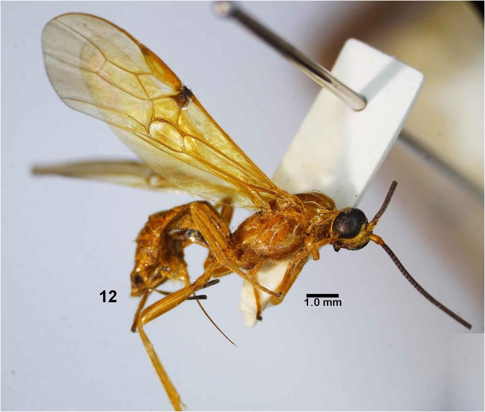
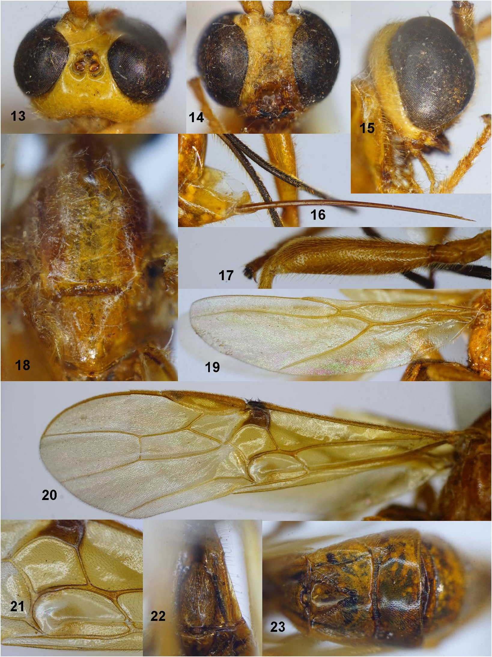
Figs 12–23 View FIGURE 12 View FIGURES 13–23
Material examined. Holotype, ♀, “Bracn. 1232 ” ( IEBR), NW Vietnam: Hoa Binh, Bao Hieu , litchi+sugarcane orchard, MT, 20°23’N 105°34’E, 80 m, 01–10.viii.2003, KD Long. GoogleMaps
Diagnosis. In frontal view, head 1.3 × as wide as long; in dorsal view, head 1.5 × as long as wide; eye length 4.3 × temple; ocelli in high triangle, POL: OD: OOL= 3: 5: 4; vein 1-M of fore wing thin, 1.6 × vein 1-CU1 ( Fig. 20 View FIGURES 13–23 ); vein 1-CU1 thick, 2.0 × as thick as vein 1-M; m-cu rather thick, slightly curved inwards, and as long as vein 1-M; basal length of second submarginal cell 3.6 × apical width; hind wing vein 1-M distinctly curved basally ( Fig. 19 View FIGURES 13–23 ); hind femur slender, 4.75 × as long as wide ( Fig. 17 View FIGURES 13–23 ); in lateral view, mesosoma 1.6 × its height; first metasomal tergite length 1.3 × its apical width; second tergite and midbasal triangular area smooth, with wide crenulated lateral convergent grooves ( Fig. 23 View FIGURES 13–23 ); antero-lateral depression convergent, rather deep, smooth; second metasomal suture finely crenulate; third tergite smooth, with fine sparse punctures; ovipositor slightly curved; sheath obliquely setose, 0.2 × as long as fore wing.
Description. Holotype, female, length of body 9.4 mm, fore wing 11.1 mm, ovipositor sheath 2.5 mm ( Fig. 12 View FIGURE 12 ).
Head. Antenna with 31 flagellomeres remaining; in lateral view, scapus 1.7 × as long as wide; length of first flagellomere 1.3 × second (9: 7); first flagellomere 1.3 × its maximum width; second flagellomere as long as wide; head 1.3 and 1.5 × as wide as long in anterior (frontal) and dorsal view respectively; face narrowed medially, rugopunctate and setose, 0.7 × as wide as long ( Fig. 14 View FIGURES 13–23 ); in frontal view, eye length 2.1 × as long as its transverse width (53: 25); clypeus separated from face with a transverse carina; height of clypeus: inter tentorial distance: tentorial ocular distance = 5: 12: 4; distance between tentorial pits 3.0 × as long as distance from tentorial pit to eye margin ( Fig. 14 View FIGURES 13–23 ); malar space 0.5 × basal width of mandible; in lateral view, transverse width of eye 5.3 × as long as temple (48: 9) ( Fig. 15 View FIGURES 13–23 ); in dorsal view, length of eye 1.7 × as long as width (39: 23), and 4.3 × temple (39: 9); ocelli in high triangle, distance between anterior and posterior ocelli 0.8 × as long as OOL; POL: OD: OOL= 3: 5: 4 ( Fig. 13 View FIGURES 13–23 ); frons flat, smooth, with fine midlongitudinal carina; vertex and temple smooth.
Mesosoma. Mesosoma 1.6 × its height (72: 44); pronotum smooth laterally, setose; mesoscutum smooth, setose, finely sparsely punctate; notauli slightly depressed anteriorly, shallow; scutellar sulcus shallow, punctate ( Fig. 18 View FIGURES 13–23 ); scutellum almost smooth, with sparse punctures; mesopleuron and metapleuron smooth, setose; medial area of metanotum without midlongitudinal carina anteriorly; propodeum smooth, setose.
Wings. Fore wing ( Fig. 20 View FIGURES 13–23 ): length of pterostigma 3.8 × its width; vein 1-SR+M curved subbasally; vein 1-M thin, 1.6 × vein 1-CU1 (21: 13); vein 1-CU1 rather thick, 2.0 × as thick as vein 1-M; vein 2-M almost straight; m-cu slightly curved inwards, and as long as vein 1-M ( Fig. 20 View FIGURES 13–23 ); subdiscal cell as broad as discal cell, with median dense setose area and narrow glabrous sclerome, and upper and posterior parts of this area glabrous ( Fig. 20 View FIGURES 13–23 ); veins SR1 curved subbasally; ratio of length of veins r: 3-RS: SR1 = 16: 41: 81; 2-RS: 3-RS: r-m = 32: 41: 19; second submarginal cell slightly narrowed apically, basal length of second submarginal cell 3.6 × apical width (75: 21); vein CU1b triangular, strongly widened basally ( Fig. 20 View FIGURES 13–23 ). Hind wing ( Fig. 19 View FIGURES 13–23 ): vein 1-M thick and strongly curved basally; vein SR slightly curved subbasally, parallel-sided apically; vein 2-SC+R vertical; subbasal cell setose; vein 1r-m almost straight; M+CU: 1-M: 1r-m = 12: 74: 28; apex of vein SC+R1 with three curved hamuli.
Legs. Fore tibia 0.8 × fore tarsus (55: 71); ratio of lengths of fore femur: tibia: basitarsus: tarsus = 47: 55: 25: 71; hind femur rather slender, 4.75 × its maximum width ( Fig. 17 View FIGURES 13–23 ); hind femur, tibia, basitarsus 4.75, 11.3 and 7.8 × their maximum width; outer and inner hind tibial spurs 0.4 × and 0.5 × as long as hind basitarsus, respectively; tarsal claw simple and with bristly ventrally.
Metasoma. First metasomal tergite 1.3 × as long as wide apically (48: 36), with a midlongitudinal carina and a pair of lateral smooth groove ( Fig. 22 View FIGURES 13–23 ); median area of first tergite striate-rugulose baso-medially, smooth coriaceous apically; second metasomal tergite 1.4 × as long as third tergite medially (28: 20), 1.4 × as wide (basally) as long (38: 28), length of midbasal triangular of second tergite in basal 0.7 × of tergite (20: 28), smooth; lateral sides of midbasal triangular area surrounded by convergent crenulate grooves ( Fig. 23 View FIGURES 13–23 ); antero-lateral depression of second tergite convergent, deep; second metasomal suture deep, crenulate; third tergite with fine dense punctures; remainder smooth; ovipositor slightly curved, needle-shaped apically, without dorsal notch or nodus and ventral serrations ( Fig. 16 View FIGURES 13–23 ); ovipositor sheath covered with dense oblique setae, 0.2 × as long as fore wing.
Colour. Body yellow; eyes dark brown; scapus and pedicellus yellow; flagellum dark brown in more than basal half, brownish yellow apically; vein and membrane of fore wing yellow, except parastigma and area between parastigma and base of vein 1-SR+M brown; all legs yellow; ovipositor sheath brown; ovipositor yellow.
Male. Unknown.
Distribution. NW Vietnam ( Hoa Binh).
Biology. Unknown.
Etymology. Named from “simulo” (Latin for “imitate, copy”), because this species is similar to M. hoabinhense , sp. nov.
Notes. Megalommum simulatum Long , sp. nov. is closely related to Megalommum hoabinhense Long , sp. nov., but can be distinguished from the latter by the following characters: 1) Vein 1-M 1.6 × vein 1-CU1 ( vs 0.9 × in M. hoabinhense ); 2) Second submarginal cell broader, parallel-sided, basal length 3.6 × its apical width ( Fig. 20 View FIGURES 13–23 ) ( vs longer and slightly narrowed apically in M. hoabinhense , 4.3 × its apical width); 3) Lateral convergent grooves of midbasal triangular area of second tergite rather wide and crenulate ( vs narrow and punctate in M. hoabinhense ).
No known copyright restrictions apply. See Agosti, D., Egloff, W., 2009. Taxonomic information exchange and copyright: the Plazi approach. BMC Research Notes 2009, 2:53 for further explanation.
|
Kingdom |
|
|
Phylum |
|
|
Class |
|
|
Order |
|
|
Family |
|
|
Genus |