
Triplocania, Roesler, 1940
|
publication ID |
https://doi.org/10.11646/zootaxa.4336.1.1 |
|
publication LSID |
lsid:zoobank.org:pub:FA65E14F-102F-4FF1-B8D5-D7E0C9126878 |
|
DOI |
https://doi.org/10.5281/zenodo.6024736 |
|
persistent identifier |
https://treatment.plazi.org/id/03DD879B-CF47-FFA8-FF6A-EA1DFA3BFEC0 |
|
treatment provided by |
Plazi |
|
scientific name |
Triplocania |
| status |
|
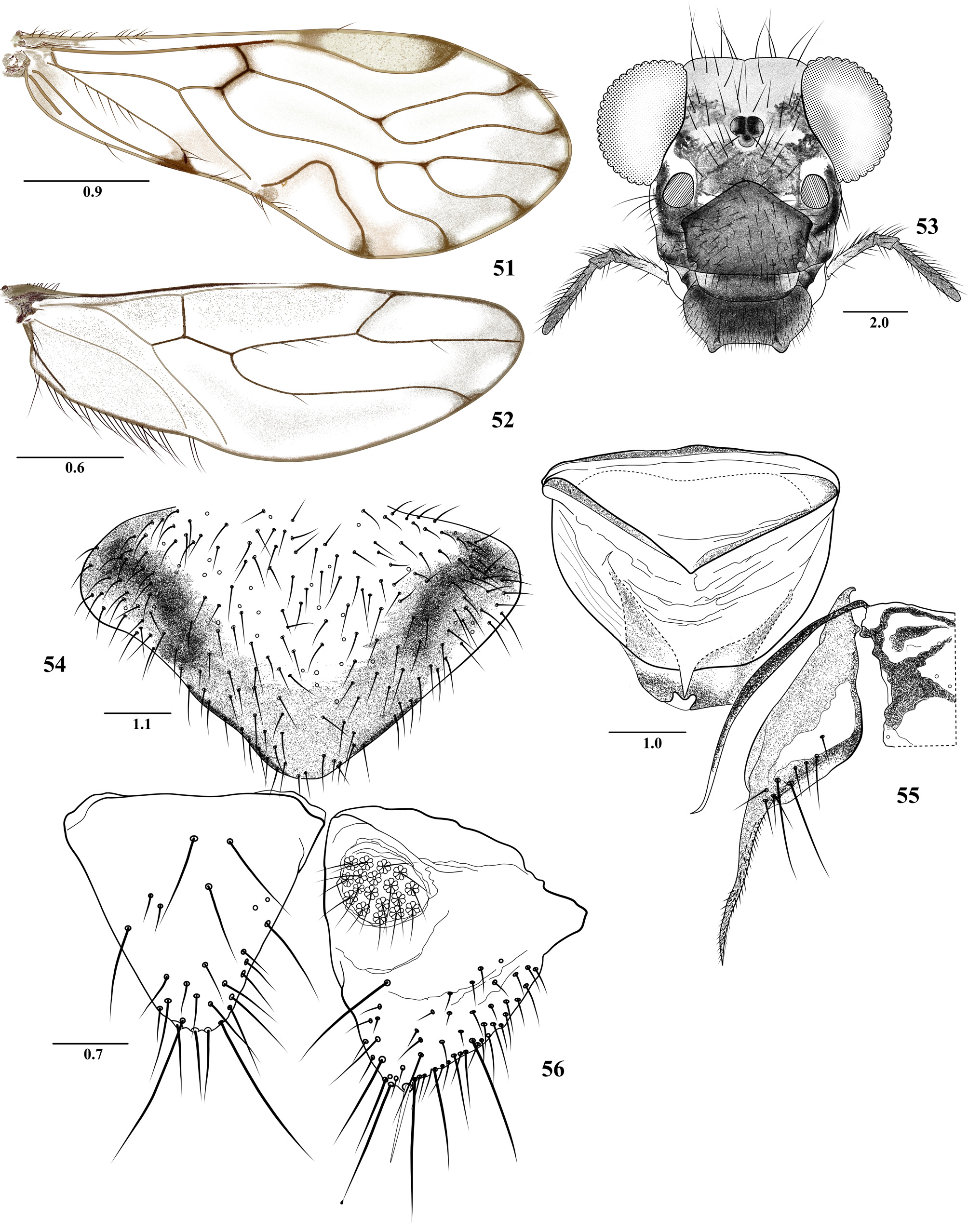
Key to the Colombian species of Triplocania View in CoL
1. Males............................................................................................... 2
- Females............................................................................................ 35
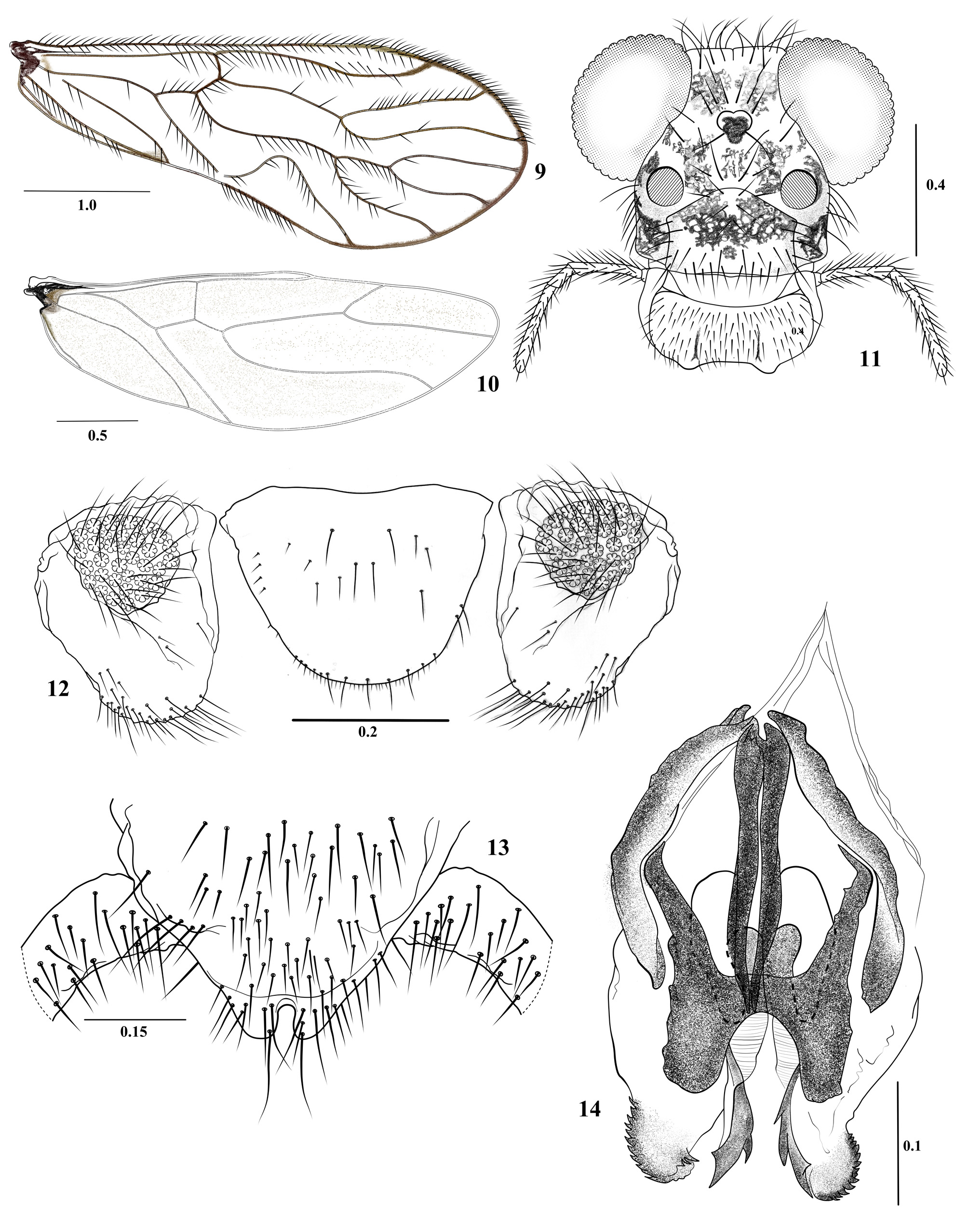
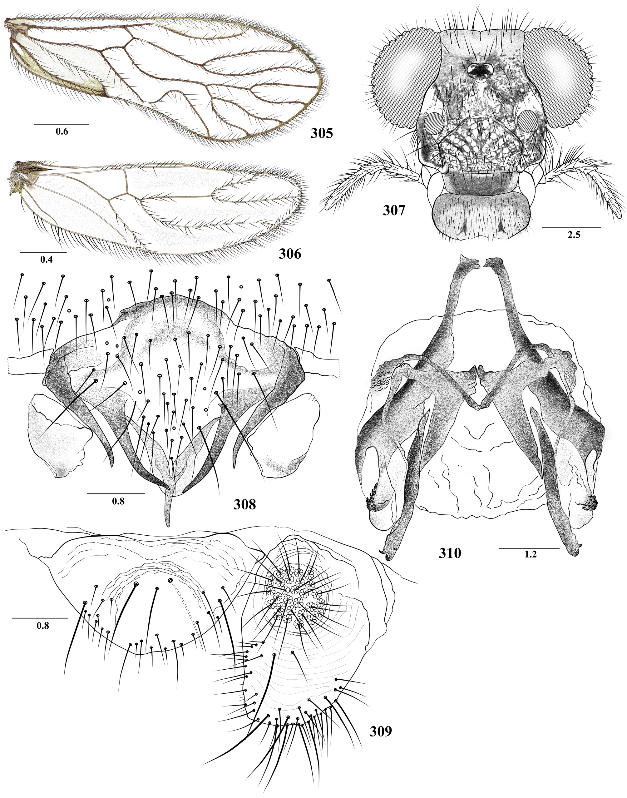
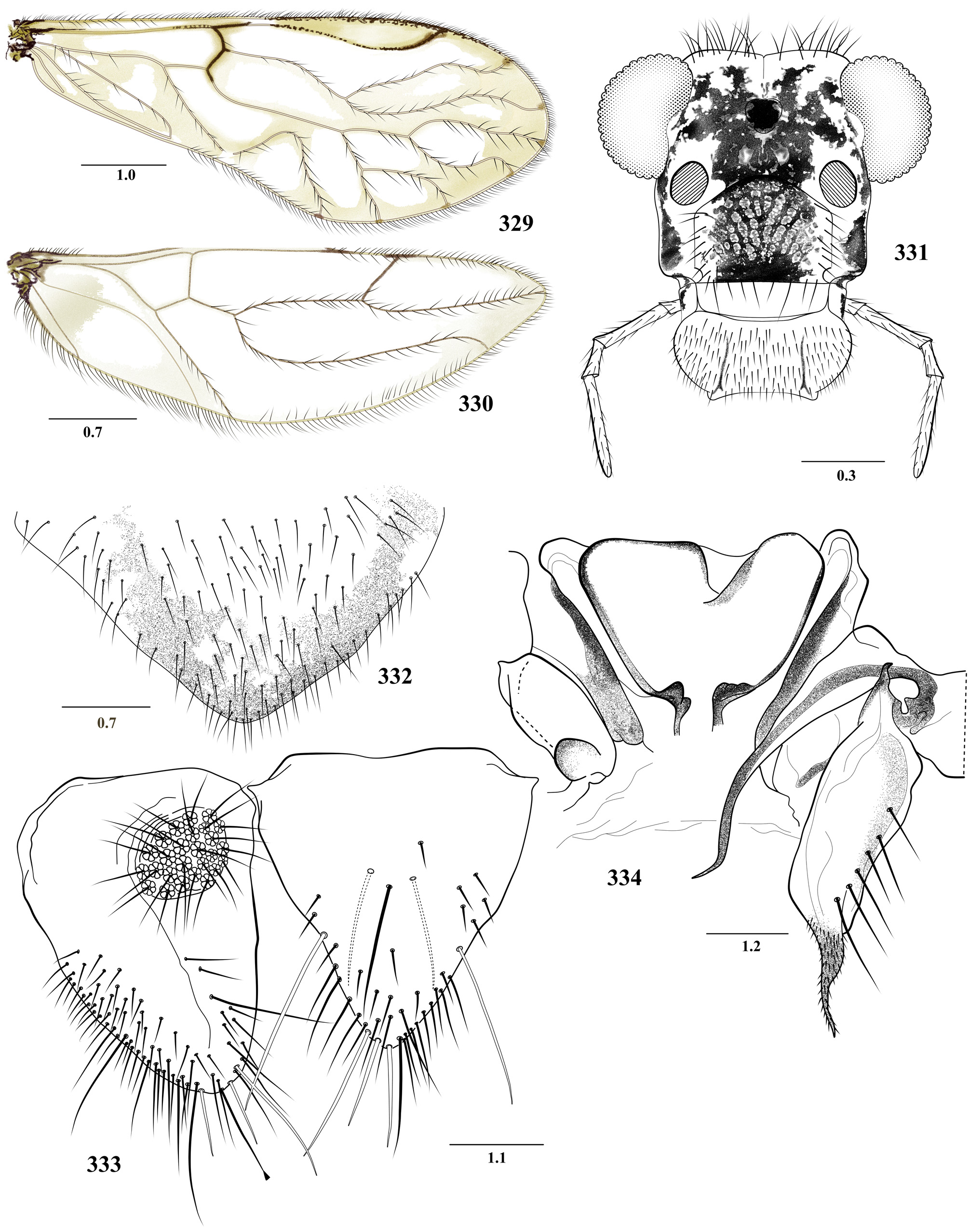
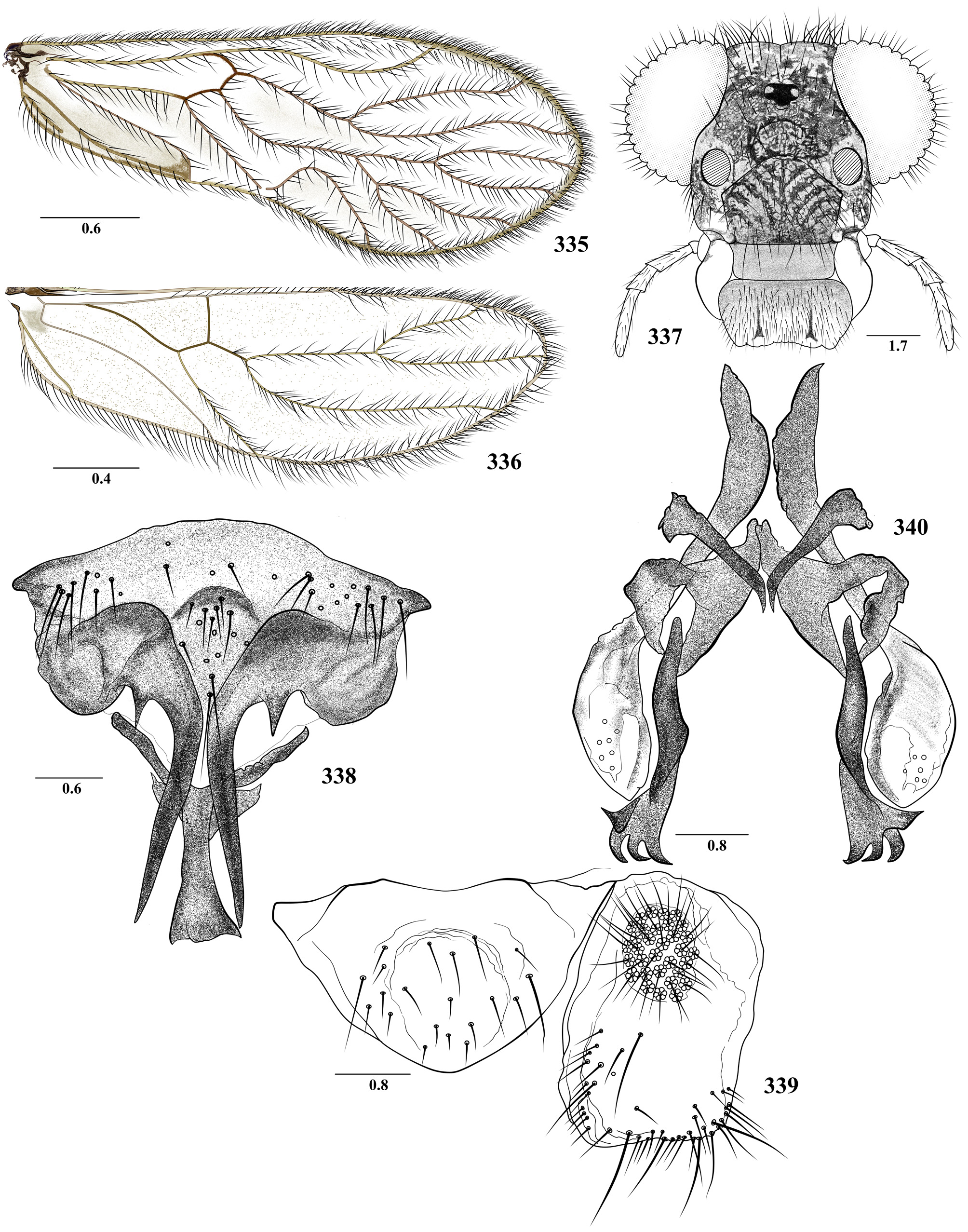
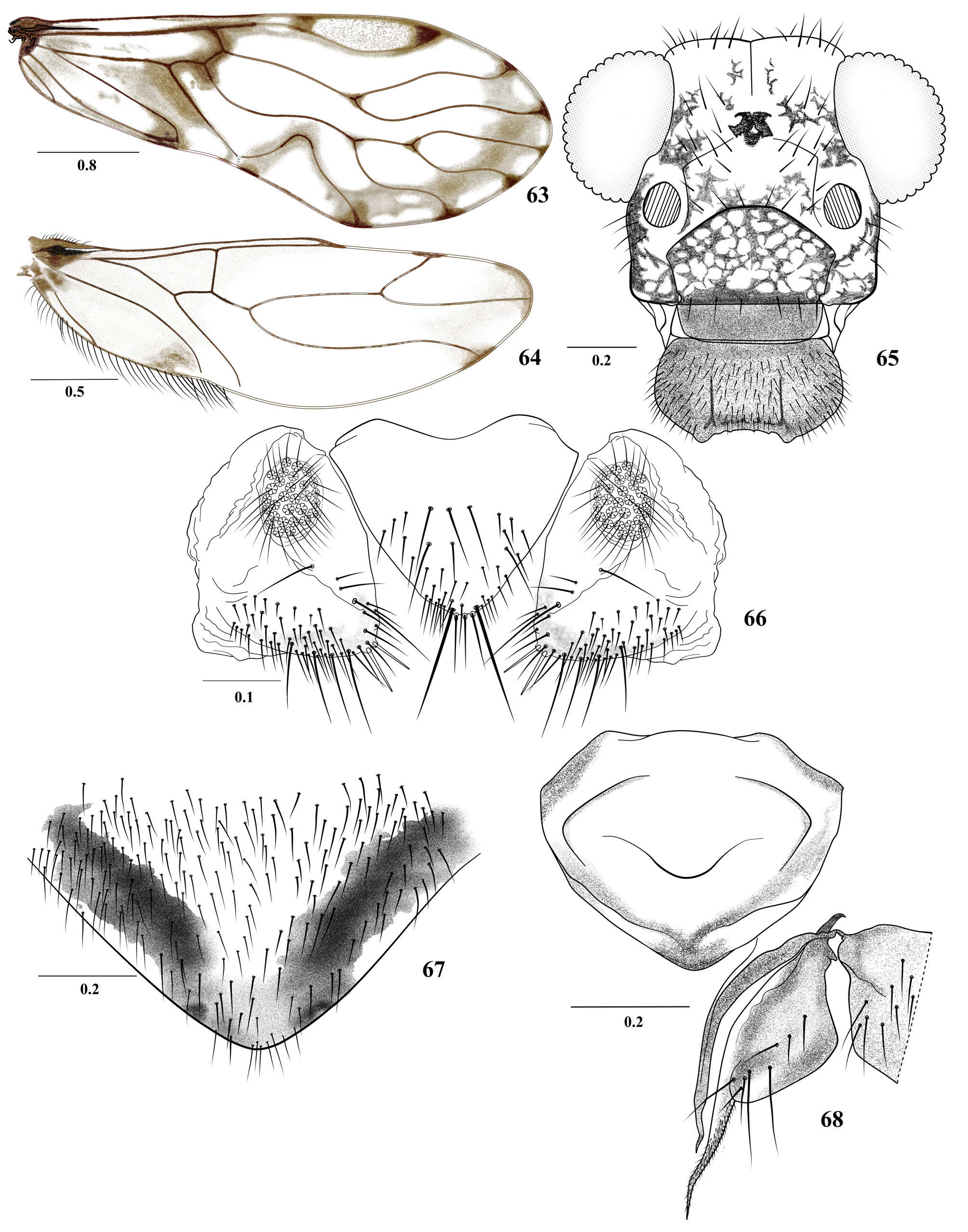
2. Forewing M3 branched ( Figs 305 View FIGURES 305 – 310 , 317 View FIGURES 317 – 322 , 323 View FIGURES 323 – 328 , 329 View FIGURES 329 – 334 , 335 View FIGURES 335 – 340 , 341 View FIGURES 341 – 346 , 347 View FIGURES 347 – 352 , 353 View FIGURES 353 – 358 and 359 View FIGURES 359 – 364 )..................................... 3
- Forewing M3 simple ( Figs 3 View FIGURES 3 – 8 , 9 View FIGURES 9 – 14 , 75 View FIGURES 75 – 80 , etc.)................................................................... 8
3. Hypandrium with only the central sclerite, this with short caudo-lateral lobular processes and one central narrow elongate process having two small apical lobes ( Fig. 326 View FIGURES 323 – 328 ); phallosome as in figure 328..................... T. lamensuraensis View in CoL n. sp.
- Hypandrium with two to four sclerites ( Figs 308 View FIGURES 305 – 310 , 338 View FIGURES 335 – 340 , 365, 365 and 367 View FIGURES 365 – 369 ); central sclerite of hypandrium and phallosome variable ( Figs 310 View FIGURES 305 – 310 , 371 View FIGURES 370 – 373 and 340 View FIGURES 335 – 340 )............................................................................. 4
4. Hypandrium of three sclerites, central sclerite with two pairs of posterior, acuminate processes, and a posterior median, elongate, acuminate process ( Figs 308 View FIGURES 305 – 310 and 365 View FIGURES 365 – 369 )................................................................. 5
- Hypandrium of two or four sclerites, the anterior-central sclerite with a pair of long acuminate, horn-shaped processes, strongly curved distally ( Figs 338 View FIGURES 335 – 340 , 366 View FIGURES 365 – 369 , and 367); posterior sclerite with an acuminate or spatulate process............... 6
5. Central sclerite of hypandrium with median, posterior, long acuminate process, almost as long as the lateral ones ( Fig. 365 View FIGURES 365 – 369 ); phallosome with phallobase almost straight proximally, posterior endophallic sclerite sinuous distally, with strongly curved tapered distal process ( Fig. 370 View FIGURES 370 – 373 )............................................................... T. furcata View in CoL New
- Central sclerite of hypandrium with median, posterior, stout process, longer than the lateral ones ( Fig. 308 View FIGURES 305 – 310 ); phallobase curved proximally toward the mesal line; posterior endophallic sclerites almost straight distally, with curved tapered apical process and two curved anteapical small teeth ( Fig. 310 View FIGURES 305 – 310 ).............................................. T. furcatoides View in CoL n. sp.
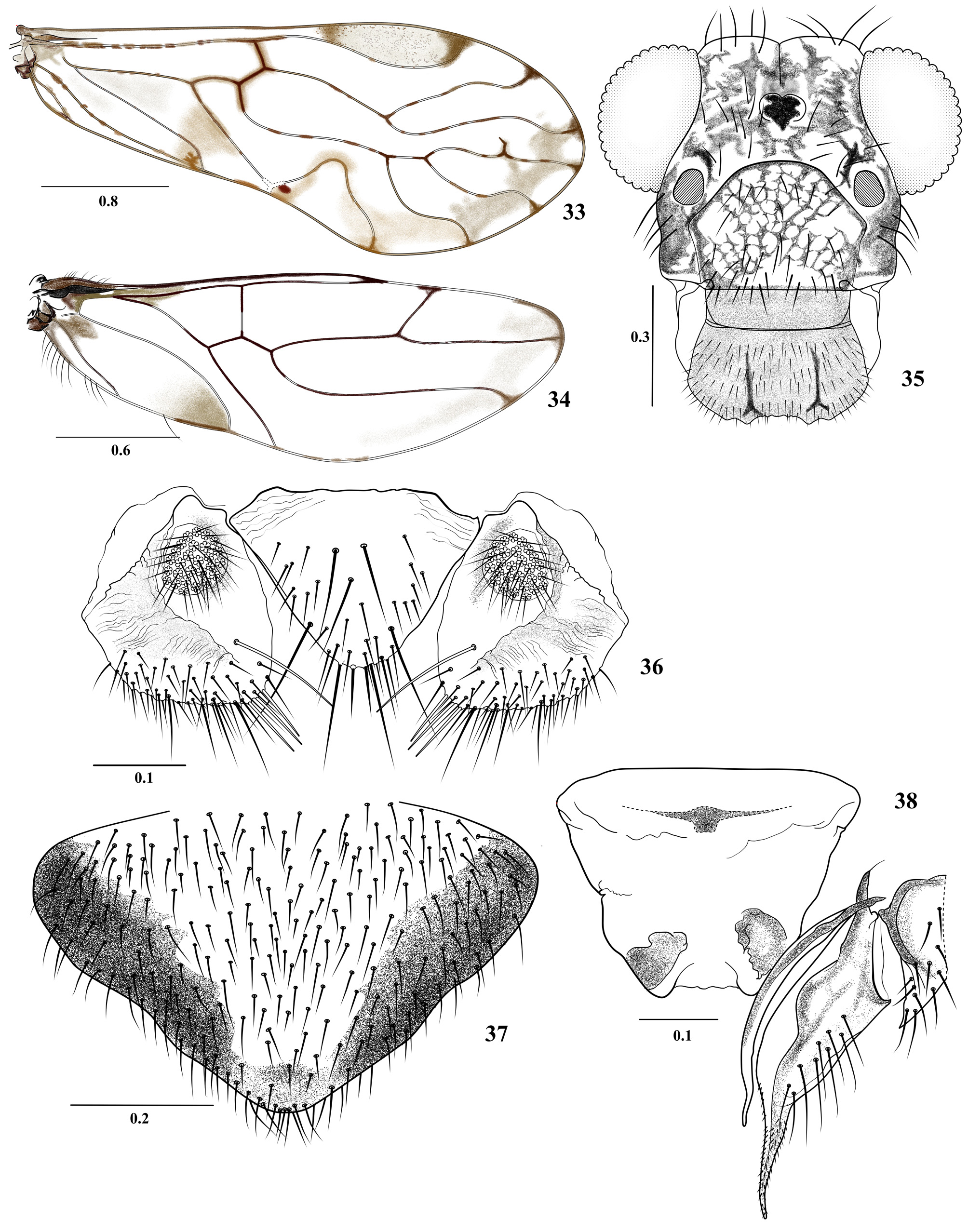
6. Hypandrium of two sclerites, acuminate processes of anterior sclerite each with a basal tooth; posterior sclerite with spatulate posterior process, dilated distally ( Fig. 338 View FIGURES 335 – 340 ); phallosome with side struts independent, each widened basally ( Fig. 340 View FIGURES 335 – 340 )........................................................................................ T. leguizamoensis View in CoL n. sp.
- Hypandrium of four sclerites, spines of anterior sclerite without basal tooth ( Fig. 366 and 367 View FIGURES 365 – 369 ); posterior sclerite with a short, stout tapered posterior process........................................................................... 7
7 Posterior sclerite of hypandrium with posterior process stout, about as long as the antero-posterior length of the anterior sclerite; sickle-shaped lateral processes of the anterior sclerite long, extending beyond the side sclerites ( Fig. 366 View FIGURES 365 – 369 )............................................................................................. T. lamasi Silva Neto et al.
- Posterior sclerite of hypandrium slender, with posterior process slender, much longer than the antero-posterior length of the anterior sclerite ( Fig. 367 View FIGURES 365 – 369 ); sickle-shaped lateral processes of the anterior sclerite short, not extending beyond the side sclerites............................................................................ T. lamasoides Silva Neto et al.
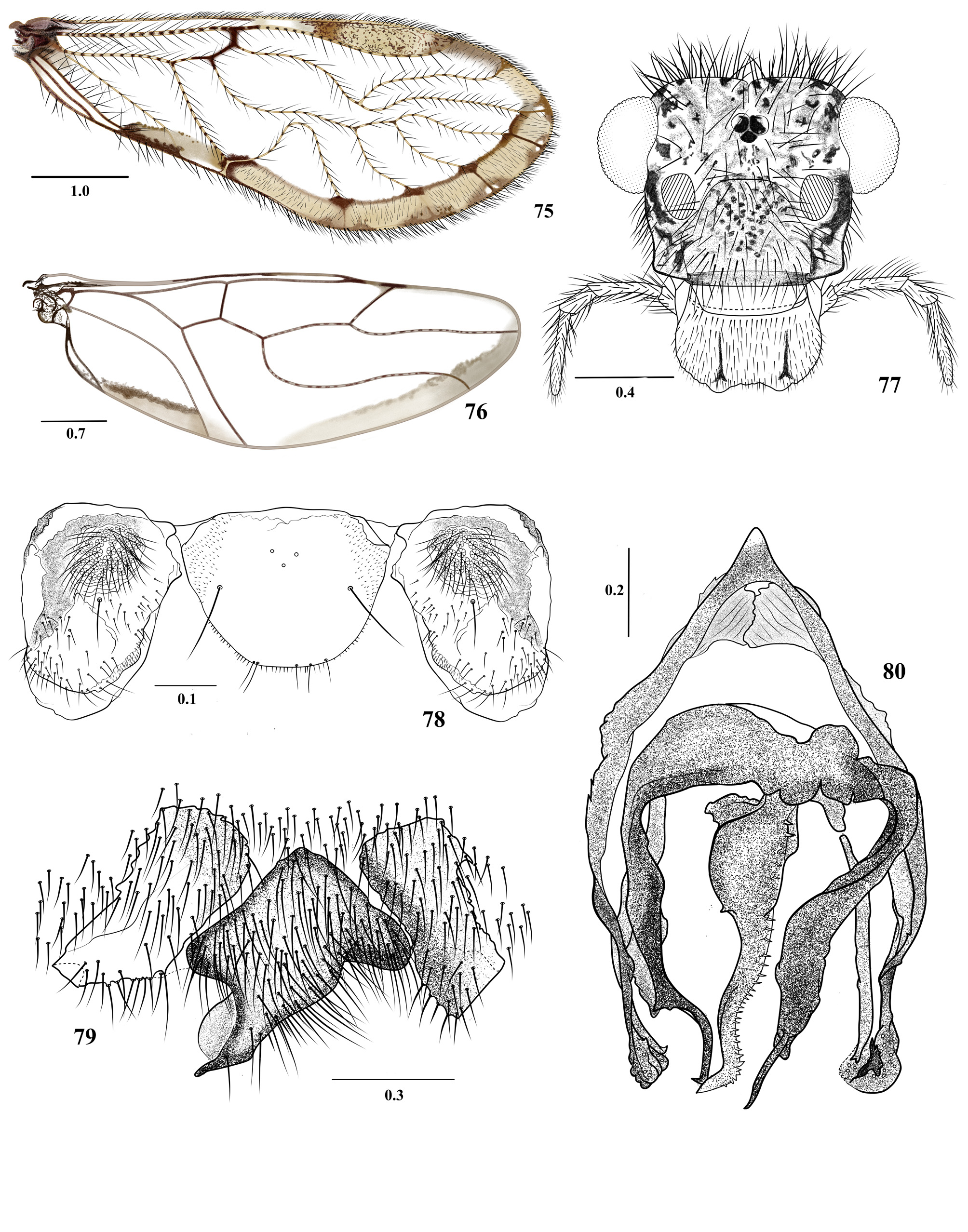
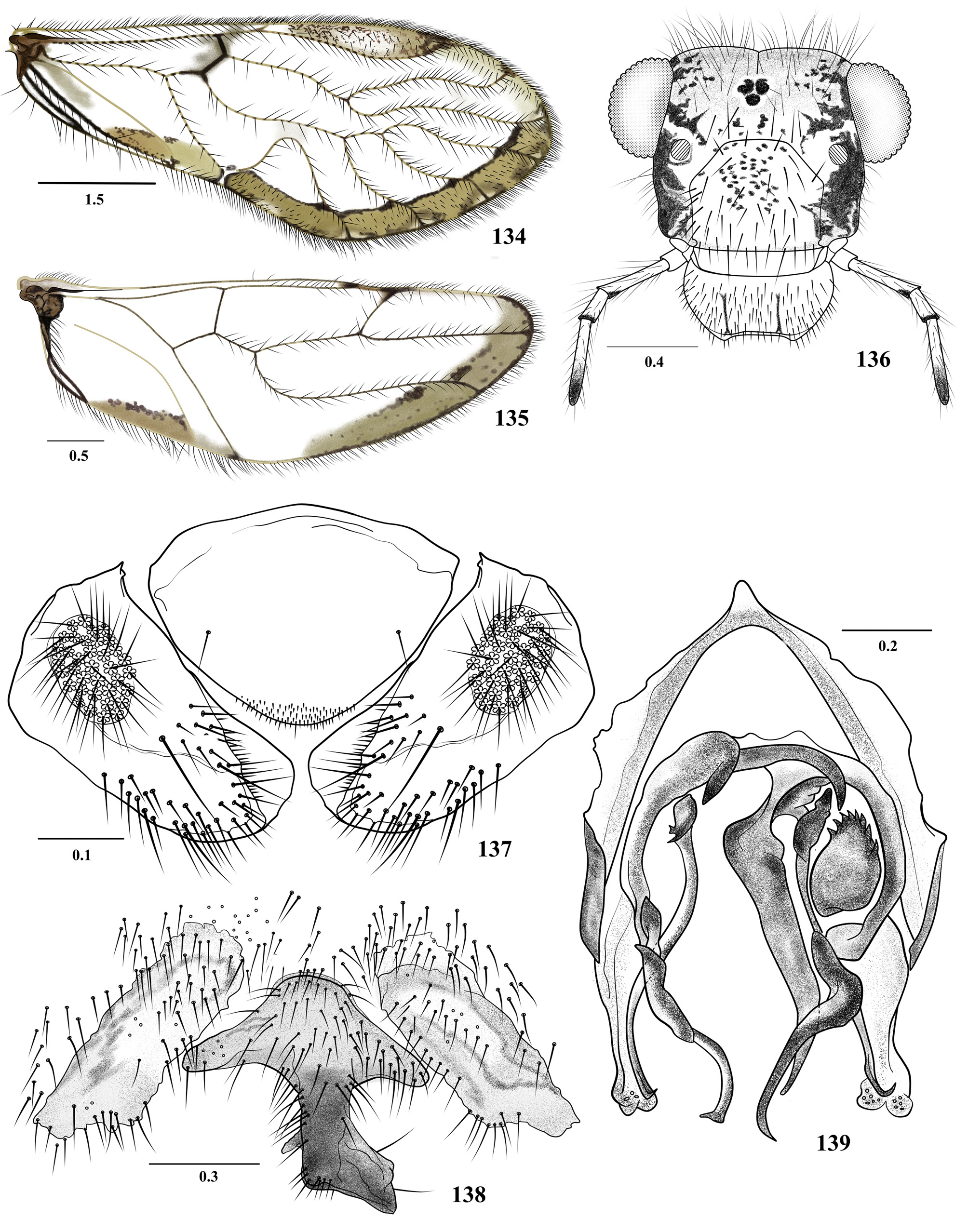
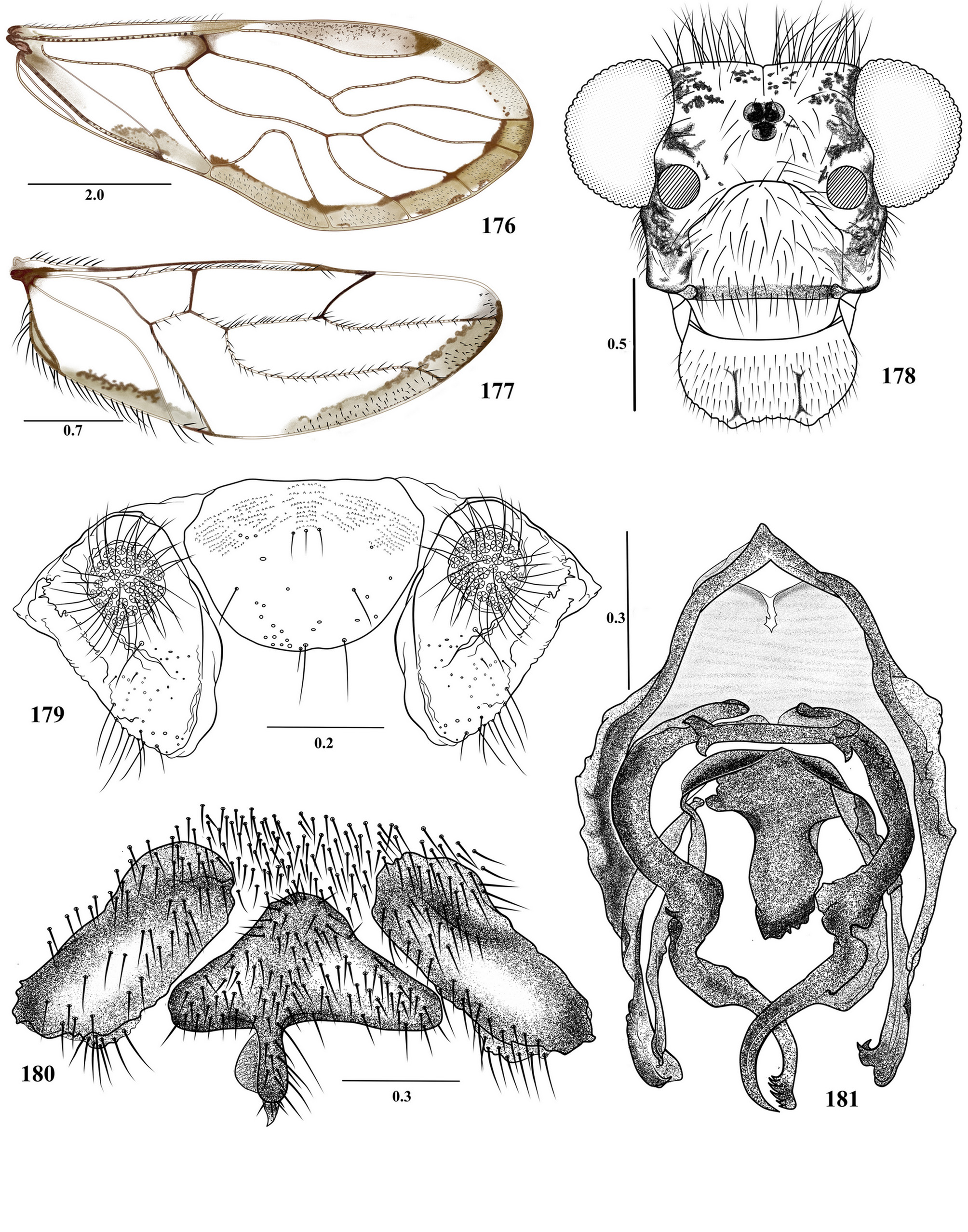
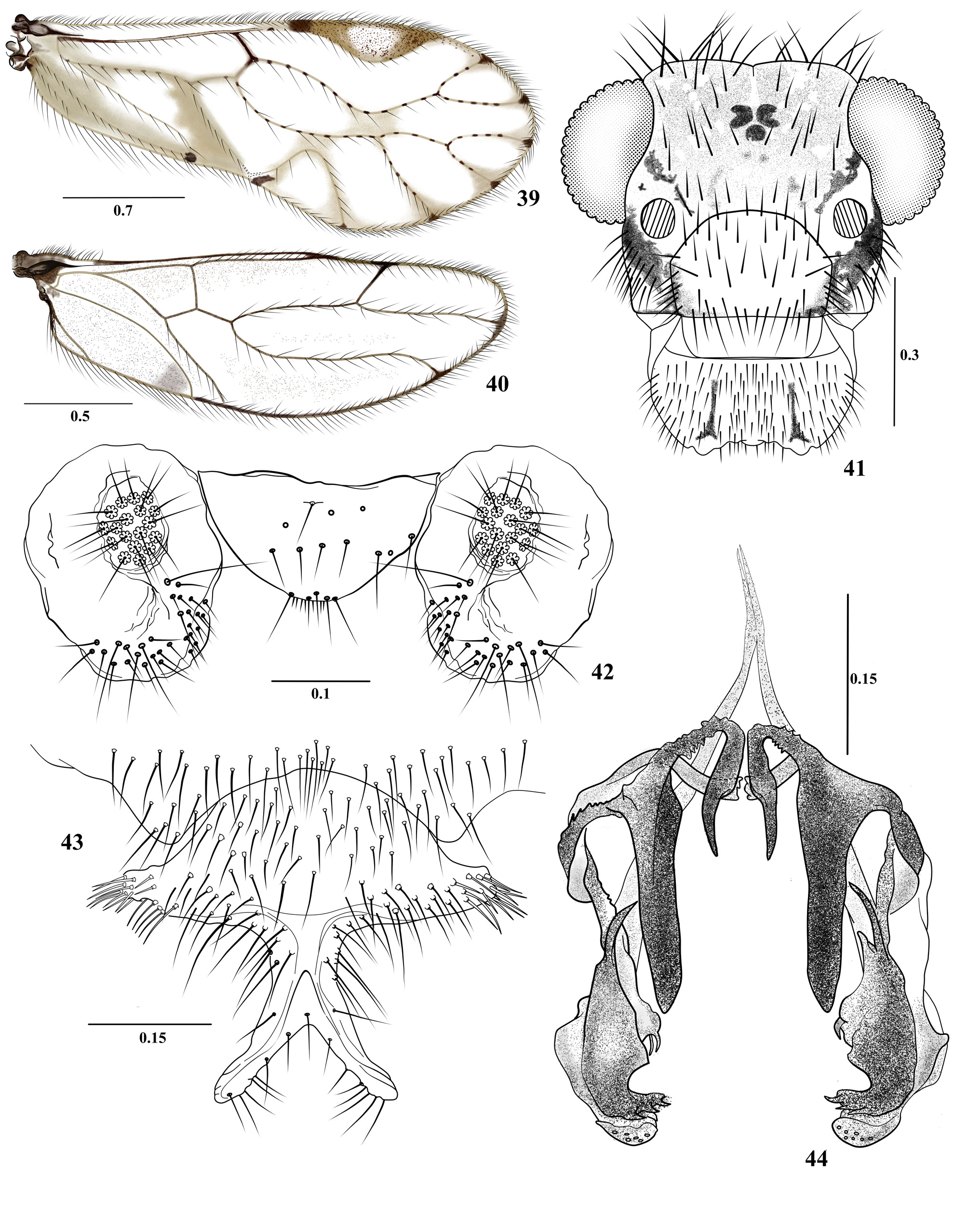
8. Central sclerite of hypandrium asymmetric, with apex directed dorsally ( Figs 79 View FIGURES 75 – 80 , 138 View FIGURES 134 – 139 , 180 View FIGURES 176 – 181 , 222 View FIGURES 218 – 223 ); forewings with a brown marginal band, from R4+5 to end of CuP; anterior endophallic sclerite of three arms or processes, one central................ 9
- Central sclerite of hypandrium symmetric ( Figs 7 View FIGURES 3 – 8 , 13 View FIGURES 9 – 14 , 18 View FIGURES 15 – 20 ), forewings hyaline or with brown marginal band; anterior endophallic sclerites paired, usually with two central processes........................................................ 12
9. Phallosome with antero-mesal endophallic sclerites projected caudally to about the level of the apex of the external parameres ( Figs 80 View FIGURES 75 – 80 and 139 View FIGURES 134 – 139 ); pair of posterior endophallic sclerites with arms not distally overlapping.......................... 10
- Phallosome with short antero-mesal endophallic sclerites, not projected caudally to the level of the apex of the external parameres ( Figs 181 View FIGURES 176 – 181 and 223 View FIGURES 218 – 223 ); pair of posterior endophallic sclerites asymmetrical, each arm anteriorly curved and distally overlapping......................................................................................... 11
10. Central sclerite of hypandrium with a stout, median posterior projection, arrow-shaped distally, slanted to the left ( Fig. 1 38 View FIGURE 1 View FIGURE 2 View FIGURES 3 – 8 View FIGURES 9 – 14 View FIGURES 15 – 20 View FIGURES 21 – 26 View FIGURES 27 – 32 View FIGURES 33 – 38 ). Endophallic sclerites slender, mesal sclerite smooth, narrowing distally, a round asymmetric sclerite anteriorly on the left, bearing a row of teeth along anterior border ( Fig. 139 View FIGURES 134 – 139 )......................................... T. felidiaensis n. sp.
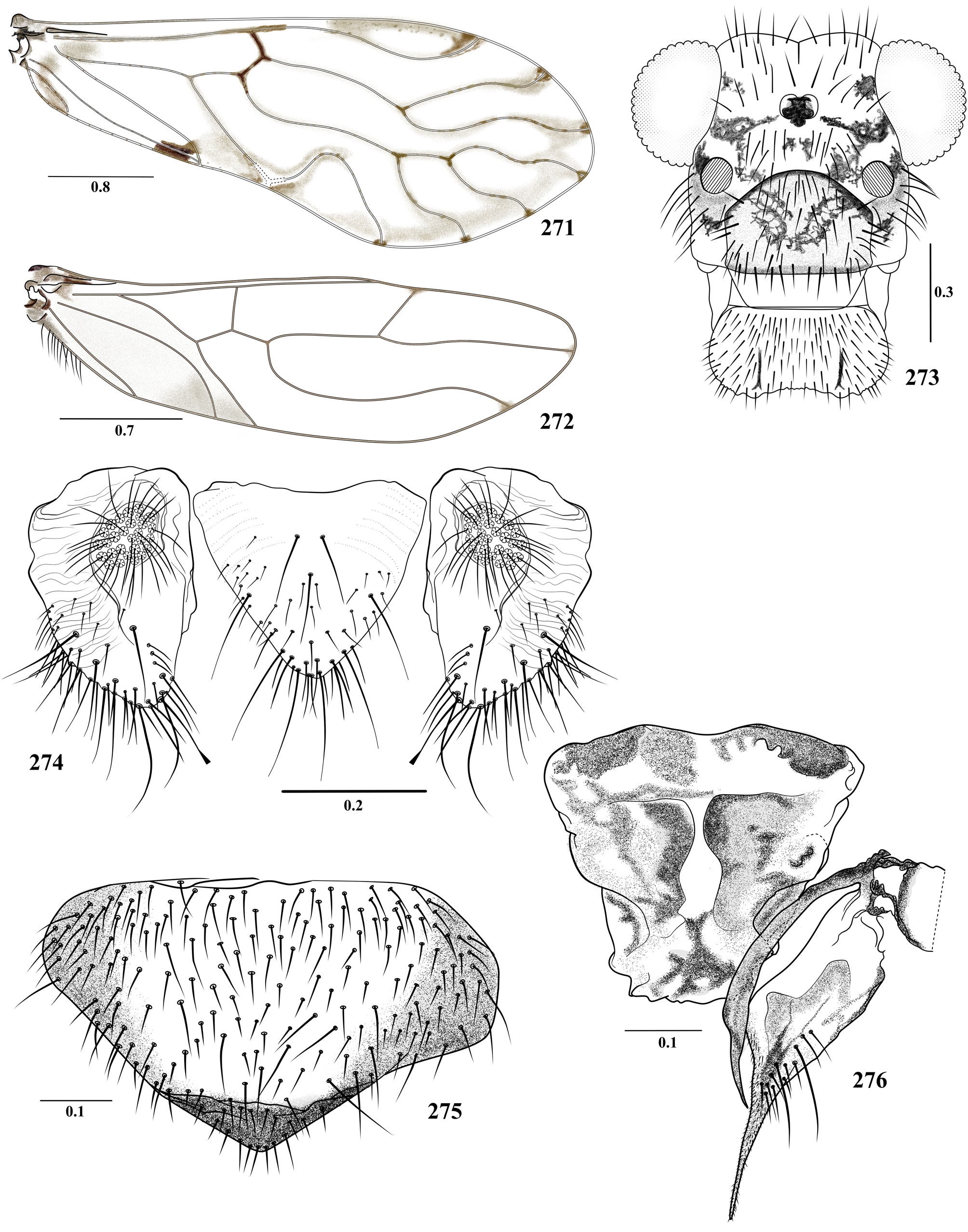
- Central sclerite of hypandrium with an acute, median posterior projection, slanted to the right ( Fig. 79 View FIGURES 75 – 80 ). Anterior pair of endophallic sclerites distally narrow, acuminate, with arms long and slender; mesal sclerite dilated proximally, narrowing distally, acuminate, bearing a row of short straight spines along side, bending to the left ( Fig. 80 View FIGURES 75 – 80 )............. T. calima n. sp.
11. Posterior pair of endophallic sclerites curved and acuminate distally; mesal sclerite with median projection stout, distally forked, with both arms acuminate, one straight and the other curved, bearing an anteapical denticle on the inner border ( Fig. 223 View FIGURES 218 – 223 )............................................................................. T. mariacarmenae n. sp.
Posterior pair of endophallic sclerites with right one distally curved, acuminate; left one distally curved, apically dilated, with a row of teeth on outer border; mesal pair with arms slender, and a stout median projection bearing distally a row of teeth ( Fig. 181 View FIGURES 176 – 181 )............................................................................... T. humboldtiana n. sp.
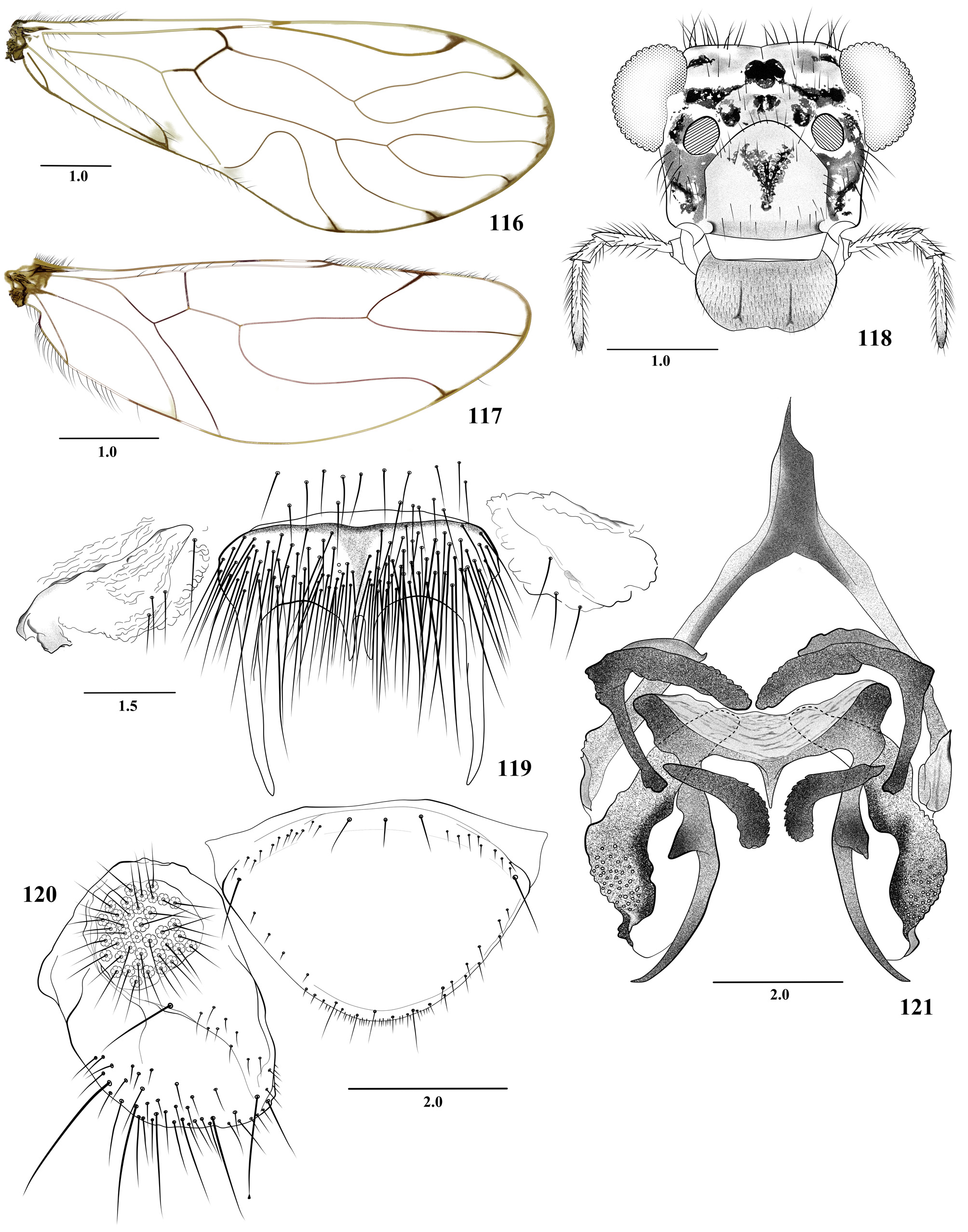
12. Central sclerite of hypandrium with a deep, U-shaped posterior concavity; postero-lateral processes elongate and tapered, widely separated; posterior endophallic sclerite tapered and curved outwards, mesal sclerite transverse ( Figs 18 View FIGURES 15 – 20 and 119 View FIGURES 116 – 121 ).. 13
- Central sclerite of hypandrium variable ( Figs 7 View FIGURES 3 – 8 , 43 View FIGURES 39 – 44 , 48 View FIGURES 45 – 50 , 61 View FIGURES 57 – 62 , 91 View FIGURES 87 – 92 , 96 View FIGURES 93 – 98 , 114 View FIGURES 110 – 115 , 150 View FIGURES 146 – 151 , 168 View FIGURES 164 – 169 , 192 View FIGURES 188 – 193 , 210 View FIGURES 206 – 211 , 234 View FIGURES 230 – 235 , 251 View FIGURES 247 – 252 , 257 View FIGURES 253 – 258 , 291 View FIGURES 287 – 292 , and 368), if Ushaped posterior concavity, then lateral processes not elongated and tapered as the previous one; endophallic sclerites variable................................................................................................... 14
13. Central sclerite of hypandrium with short acuminate teeth proximally on the inner side ( Fig. 18 View FIGURES 15 – 20 ); posterior endophallic sclerite strongly enlarged basally and with short process outwards; mesal sclerite transverse ( Fig. 20 View FIGURES 15 – 20 )............ T. andaqui n. sp.
- Central sclerite of hypandrium with two teeth or mesal processes ( Fig. 119 View FIGURES 116 – 121 ); posterior endophallic sclerite enlarged basally but not as the previous one ( Fig. 121 View FIGURES 116 – 121 )............................................................. T. dimitrii n. sp.
14. Vein A2 not reaching the wing margin.................................................................... 15
- Vein A2 reaching the wing margin....................................................................... 30
15. Central sclerite of hypandrium with caudo-lateral and central projections ( Figs 7 View FIGURES 3 – 8 , 96 View FIGURES 93 – 98 , 150 View FIGURES 146 – 151 , 257 View FIGURES 253 – 258 and 291 View FIGURES 287 – 292 )................ 16
- Central sclerite of hypandrium with caudo-lateral projections only, widely separated or close and centered, or reduced to a single mesal lobe ( Figs 43 View FIGURES 39 – 44 , 48 View FIGURES 45 – 50 , 61 View FIGURES 57 – 62 , 91 View FIGURES 87 – 92 , 114 View FIGURES 110 – 115 , 168 View FIGURES 164 – 169 , 192 View FIGURES 188 – 193 , 210 View FIGURES 206 – 211 , 234 View FIGURES 230 – 235 , 251 View FIGURES 247 – 252 and 368 View FIGURES 365 – 369 )........................................ 20
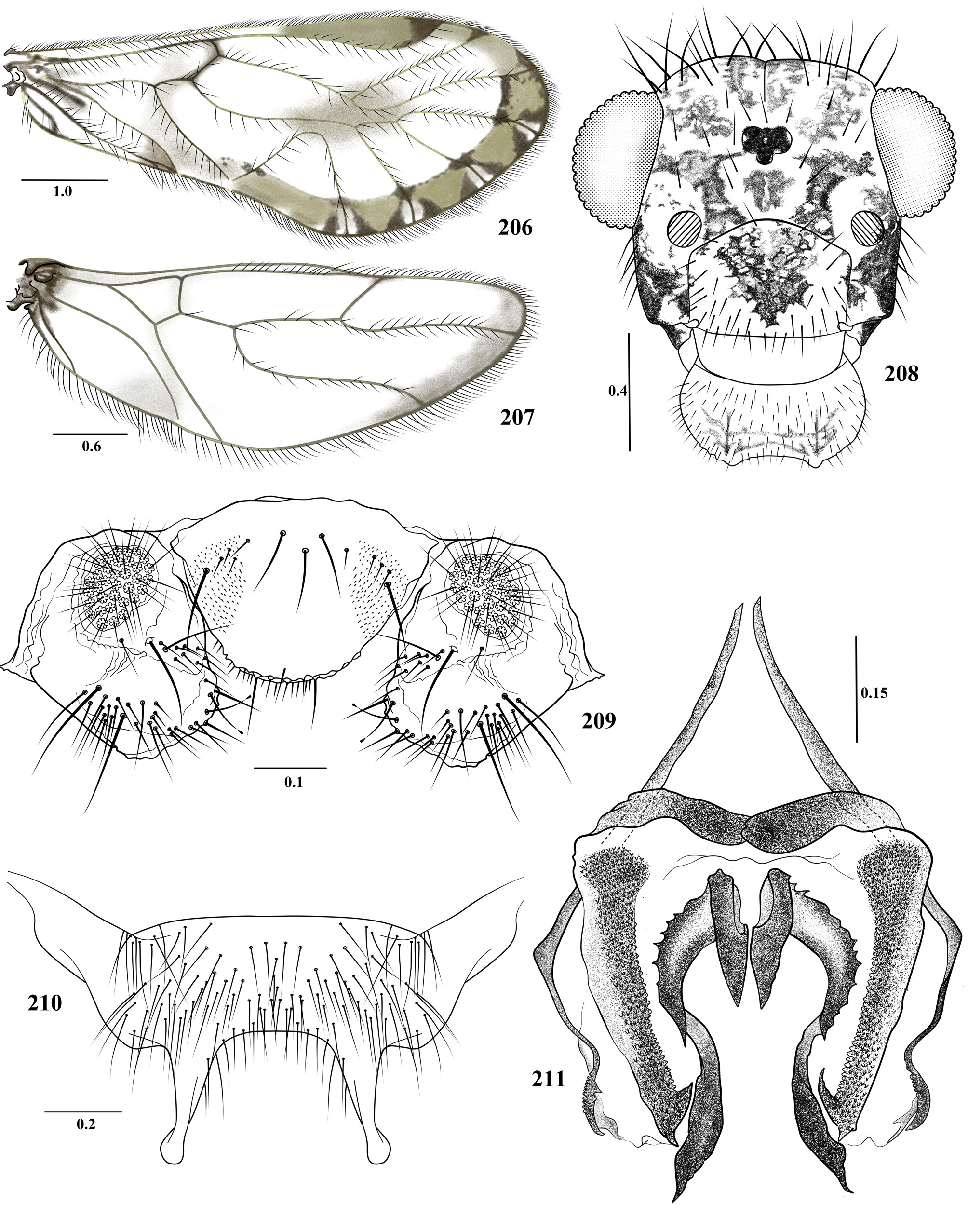
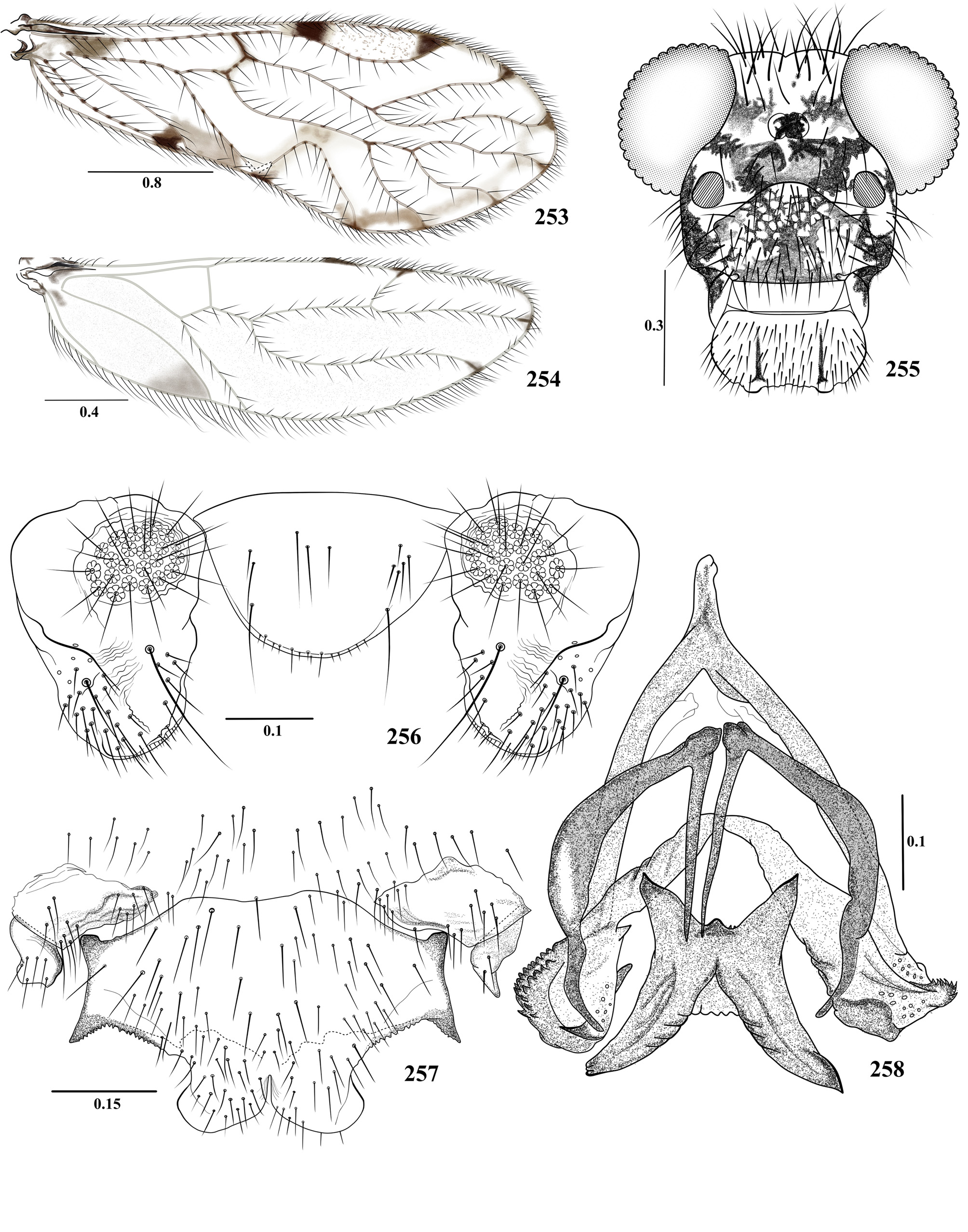
16. Central sclerite of hypandrium on inner margin of lateral processes and between these and median lobes with short sclerotized teeth; median lobes wide, occupying more than 1/3 of the posterior margin, distally rounded and directed laterally ( Fig. 257 View FIGURES 253 – 258 ); phallosome with mesal endophallic sclerite stout, X-shaped, anterior sclerites with inner arms straight, slender, acuminate, and outer arms stout, bow shaped ( Fig. 258 View FIGURES 253 – 258 ).................................................... T. pericosensis n. sp.
- Central sclerite of hypandrium without teeth as described above, median lobes not as wide; phallosome with mesal sclerite Wshaped ( Fig. 96 View FIGURES 93 – 98 ) or as two separate sclerites ( Fig. 8 View FIGURES 3 – 8 )......................................................... 17
17. External parameres distally rounded, directed posteriorly; mesal sclerites laminar, proximally wide, narrowing distally, with apical teeth; anterior sclerites with inner arms globose, outer arms curved, distally blunt ( Figs. 8 View FIGURES 3 – 8 )... T. amacayacuensis n. sp.
- External parameres not as above; mesal sclerite W-shaped ( Fig. 96 View FIGURES 93 – 98 )............................................. 18
18. Central sclerite of hypandrium with well developed postero-lateral processes ( Figs 150 View FIGURES 146 – 151 , 291 View FIGURES 287 – 292 )........................ 19
- Central sclerite of hypandrium with postero-lateral processes small or not evident ( Fig. 96 View FIGURES 93 – 98 ); endophallic sclerites as in figure 98..................................................................................... T. cantatis n. sp.
19. Central sclerite of hypandrium with inwards curved postero-lateral processes, with stout, deeply obtusely concave median Ushaped process ( Fig. 291 View FIGURES 287 – 292 ); anterior endophallic sclerites with inner arms stout, straight, outer arms pedunculate, distally dilated, papillose ( Fig. 292 View FIGURES 287 – 292 )................................................................ T. yanacona n. sp.
- Central sclerite of hypandrium with outwardly directed postero-lateral processes, with stout, deeply obtusely concave median process, with setose lobes ( Fig. 150 View FIGURES 146 – 151 ); phallosome without anterior endophallic sclerite projected laterally and enlarged distal area ( Fig.151 View FIGURES 146 – 151 )..................................................................... T. garciamarquezi n. sp.
20. Forewings with broad marginal band from R2+3 to Cu1a ( Fig. 206 View FIGURES 206 – 211 ). Postero-lateral processes of central sclerite of hypandrium distally bulged ( Fig. 210 View FIGURES 206 – 211 ); anterior endophallic sclerite with arms L-shaped, the longer stem projected posteriorly, ending in a short process and bearing a large subapical tooth on inner border; mesal pair with outer arm curved, distally acuminate, outer border with a row of small pointed processes; inner arm straight, distally acuminate; posterior pair with arms curved, elongate, distally acuminate ( Fig. 211 View FIGURES 206 – 211 )............................................................... T. lithophila n. sp.
- Forewings without marginal pigmented band, or, if present, poorly defined ( Fig. 39 View FIGURES 39 – 44 ); postero-lateral processes of central sclerite of hypandrium variable ( Figs 43 View FIGURES 39 – 44 , 48 View FIGURES 45 – 50 , 61 View FIGURES 57 – 62 , 91 View FIGURES 87 – 92 , 114 View FIGURES 110 – 115 , 168 View FIGURES 164 – 169 , 192 View FIGURES 188 – 193 , 234 View FIGURES 230 – 235 , 251 View FIGURES 247 – 252 and 368 View FIGURES 365 – 369 ); mesal endophallic sclerite, H, W, X, or Yshaped............................................................................................. 21
21. Central sclerite of hypandrium elongate, with outer subparallel borders that continue in two processes or elongate lateral lobes widely separated or relatively close ( Figs 61 View FIGURES 57 – 62 and 168 View FIGURES 164 – 169 )........................................................ 22
- Central sclerite of hypandrium short and narrow or wider than long, with rounded or irregular outer borders ( Figs 43 View FIGURES 39 – 44 , 48 View FIGURES 45 – 50 , 91 View FIGURES 87 – 92 , 114 View FIGURES 110 – 115 , 192 View FIGURES 188 – 193 , 234 View FIGURES 230 – 235 , 251 View FIGURES 247 – 252 and 368 View FIGURES 365 – 369 )............................................................................ 23
22. Central sclerite of hypandrium with sides almost parallel, lateral processes widely separated, rounded, each bearing distally two lateral acuminate processes, and a small acuminate process on inner border ( Fig. 61 View FIGURES 57 – 62 ); endophallic sclerites as in figure 62...................................................................................... T. bicornuta n. sp.
- Central sclerite of hypandrium with sides convergent, lateral processes blunt ended; endophallic sclerites as in figure 169............................................................................................ T. huitota n. sp.
23. Hypandrium of one sclerite, wider than long, with a slender, sclerotized process in the middle ( Fig. 48 View FIGURES 45 – 50 ); anterior endophallic sclerite transverse, sausage-shaped, with a row of short denticles along posterior border; mesal sclerite broadly H-shaped, with anterior arms stout; posterior pair with each arm of two pieces, the anterior one wide, anteriorly acuminate, the posterior one elongate, curved, distally wider, with denticles along posterior border ( Fig. 50 View FIGURES 45 – 50 )............................ T. awa n. sp.
- Hypandrium of one or three sclerites; central sclerite with double processes, central or lateral ( Figs 43 View FIGURES 39 – 44 , 91 View FIGURES 87 – 92 , 114 View FIGURES 110 – 115 , 192 View FIGURES 188 – 193 , 234 View FIGURES 230 – 235 , 251 View FIGURES 247 – 252 and 368 View FIGURES 365 – 369 ); endophallic sclerites not as above................................................................ 24
24. Central sclerite of hypandrium with central short or elongate lobular projections ( Figs 43 View FIGURES 39 – 44 , 114 View FIGURES 110 – 115 , 192 View FIGURES 188 – 193 and 368 View FIGURES 365 – 369 )............ 25
- Central sclerite of hypandrium with only lobed lateral projections ( Figs 91 View FIGURES 87 – 92 , 234 View FIGURES 230 – 235 and 251 View FIGURES 247 – 252 )............................ 28
25. Central sclerite of hypandrium with long posterior process in the middle, deeply obtusely concave distally ( Fig. 43 View FIGURES 39 – 44 ); anterior endophallic sclerite small, transverse, close together in the middle, mesal pair with each arm T-shaped, with a median stout process flanked by small slender processes formed by two lateral sclerites; posterior pair proximally slender, acuminate, wider in the middle, distally curved inward, with the border denticulate ( Fig. 44 View FIGURES 39 – 44 )........................... T. asisensis n. sp.
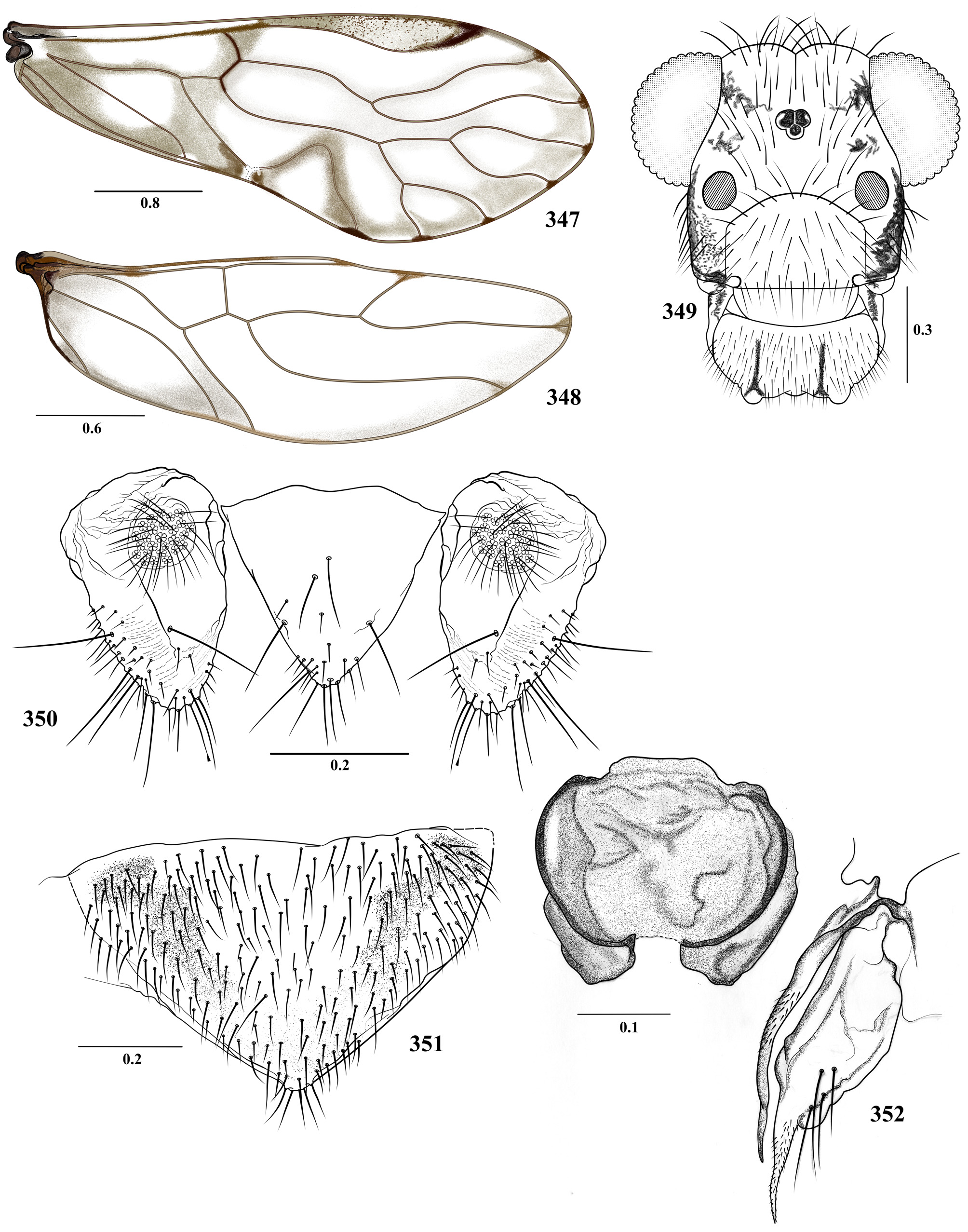
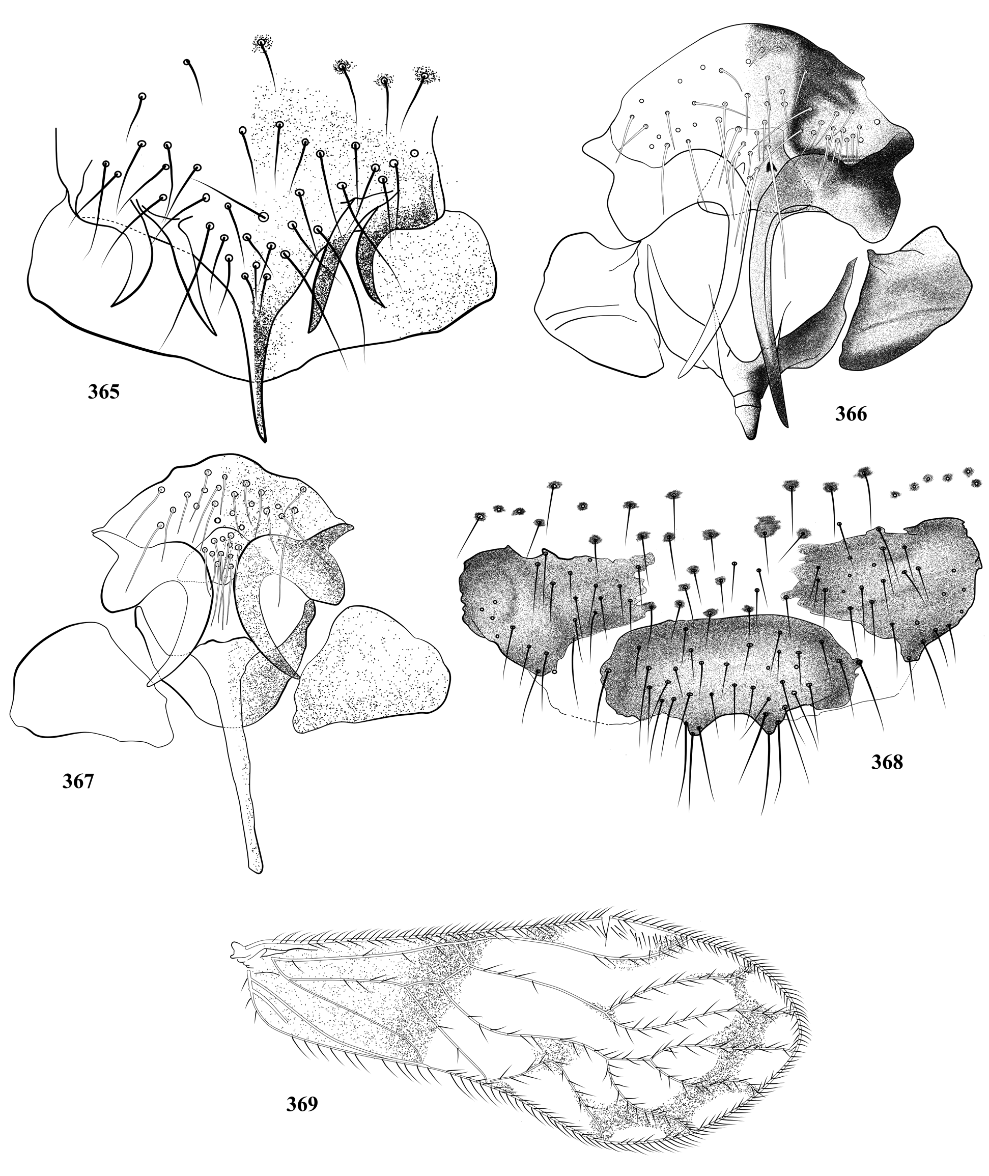
- Central sclerite of hypandrium with short lobular processes, originated in a mesal lobe wider than long ( Figs 114 View FIGURES 110 – 115 , 192 View FIGURES 188 – 193 and 368 View FIGURES 365 – 369 ); endophallic sclerites not as above ( Figs 115 View FIGURES 110 – 115 , 193 View FIGURES 188 – 193 and 371 View FIGURES 370 – 373 )................................................ 26
26. Central sclerite of hypandrium with two small median processes, almost closing a round concavity, sclerotized along its border ( Fig. 114 View FIGURES 110 – 115 ); anterior endophallic sclerites M-shaped, stout; mesal sclerite broadly X-shaped, underlaid by elliptic round areas; posterior pair slender, curved outwards, bearing denticles on outer border ( Fig. 115 View FIGURES 110 – 115 ).................. T. chocoensis n. sp.
- Central sclerite of hypandrium uniformly sclerotized, wider than long, with short lobular projections, originating in a mesal lobe wider than long ( Figs 192 View FIGURES 188 – 193 and 368 View FIGURES 365 – 369 )................................................................... 27
27. Central sclerite of hypandrium with a median, bilobed posterior process, postero-lateral corners not projected ( Fig. 192 View FIGURES 188 – 193 ); posterior endophallic sclerites of two symmetrical T-shaped arms ( Fig. 193 View FIGURES 188 – 193 ).............................. T. kichwa n. sp.
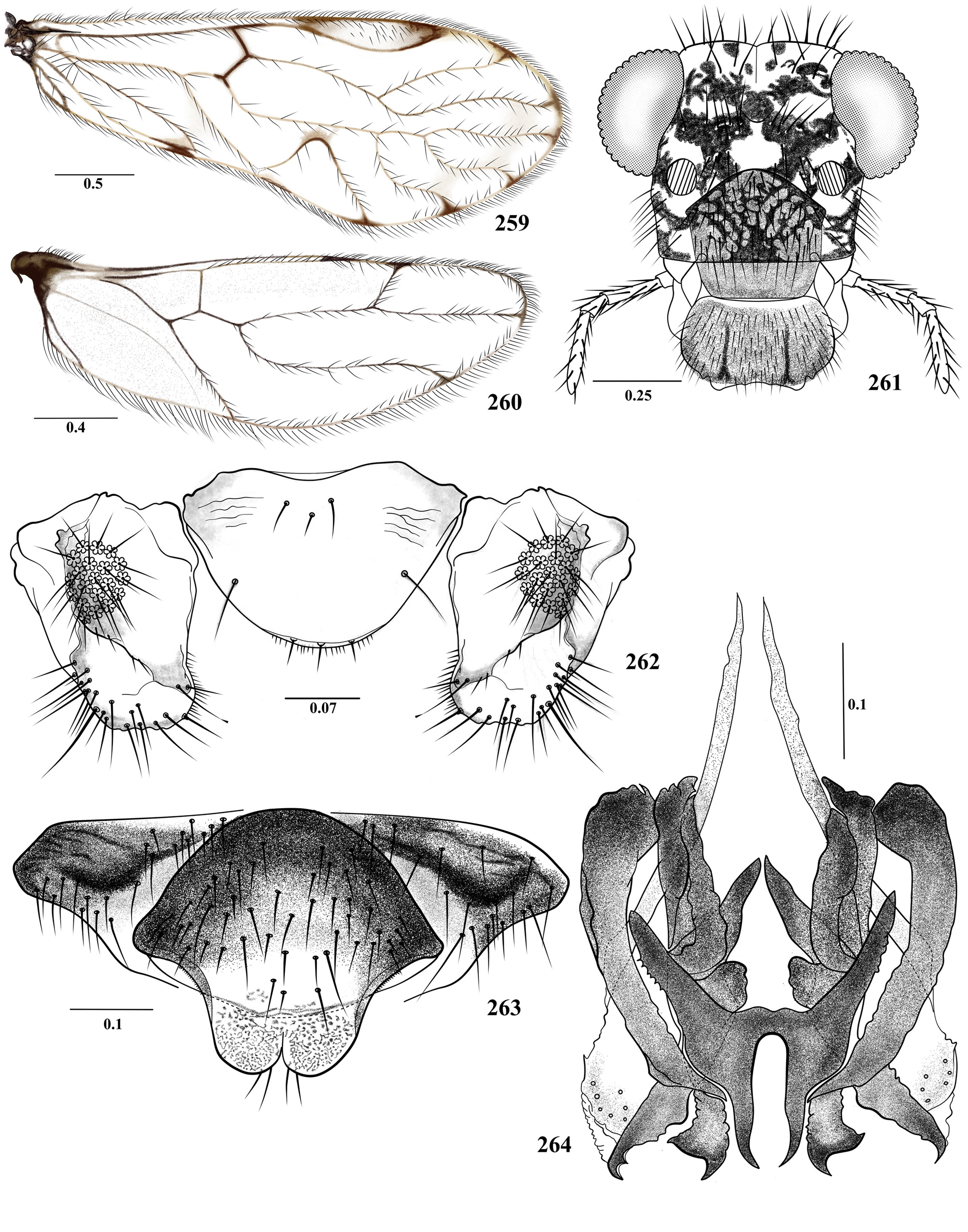
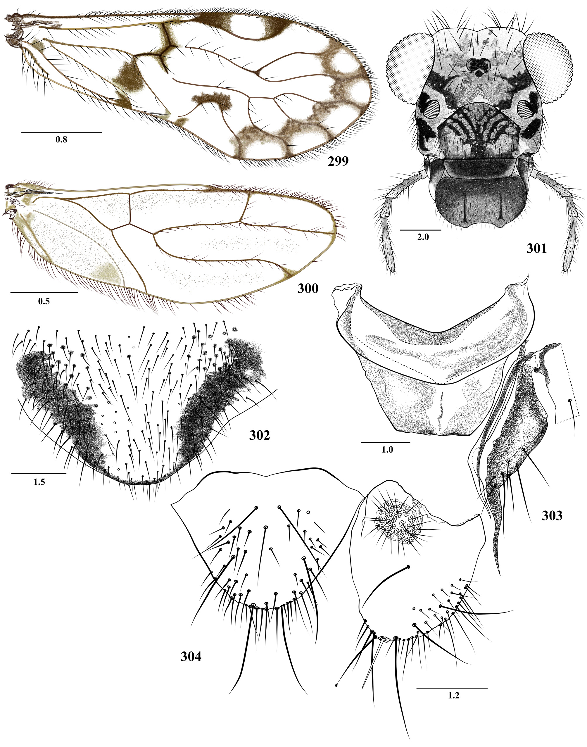
- Central sclerite of hypandrium with two small, median posterior processes, postero-lateral corners projected ( Fig. 368 View FIGURES 365 – 369 ); posterior endophallic sclerites triangular ( Fig. 371 View FIGURES 370 – 373 )........................................... T. erwini Silva-Neto et al.
28. Central sclerite of hypandrium U-shaped, stout ( Fig. 251 View FIGURES 247 – 252 ); mesal endophallic sclerite wide, H-shaped ( Fig. 252 View FIGURES 247 – 252 )................................................................................................... T. panchei n. sp.
- Central sclerite of hypandrium not as above ( Figs 91 View FIGURES 87 – 92 and 234 View FIGURES 230 – 235 ); mesal endophallic sclerite wide, H or sea star-shaped ( Figs 92 View FIGURES 87 – 92 and 235 View FIGURES 230 – 235 )............................................................................................ 29
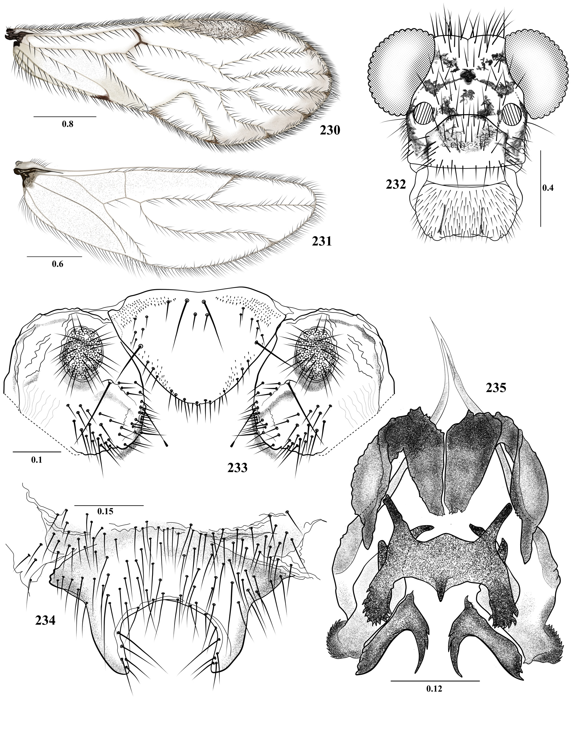
29. Central sclerite of hypandrium with two elongate postero-lateral lobes, slightly curved inwards, blunt ended ( Fig. 234 View FIGURES 230 – 235 ); mesal endophallic sclerite broadly H-shaped, posterior arms distally denticulate; anterior pair robust, U-shaped ( Fig. 235 View FIGURES 230 – 235 ).............................................................................................. T. mocoaensis n. sp.
- Central sclerite of hypandrium with two stout, distally truncate postero-lateral lobes ( Fig. 91 View FIGURES 87 – 92 ); mesal endophallic sclerite starshaped ( Fig. 92 View FIGURES 87 – 92 )......................................................................... T. camentsa n. sp.
30. Central sclerite of hypandrium with central and lateral processes ( Figs 73 View FIGURES 69 – 74 and 269 View FIGURES 265 – 270 )................................ 31
- Central sclerite of hypandrium with lateral or central processes ( Figs 13 View FIGURES 9 – 14 , 31 View FIGURES 27 – 32 , 132 View FIGURES 128 – 133 and 263 View FIGURES 259 – 264 )........................... 32
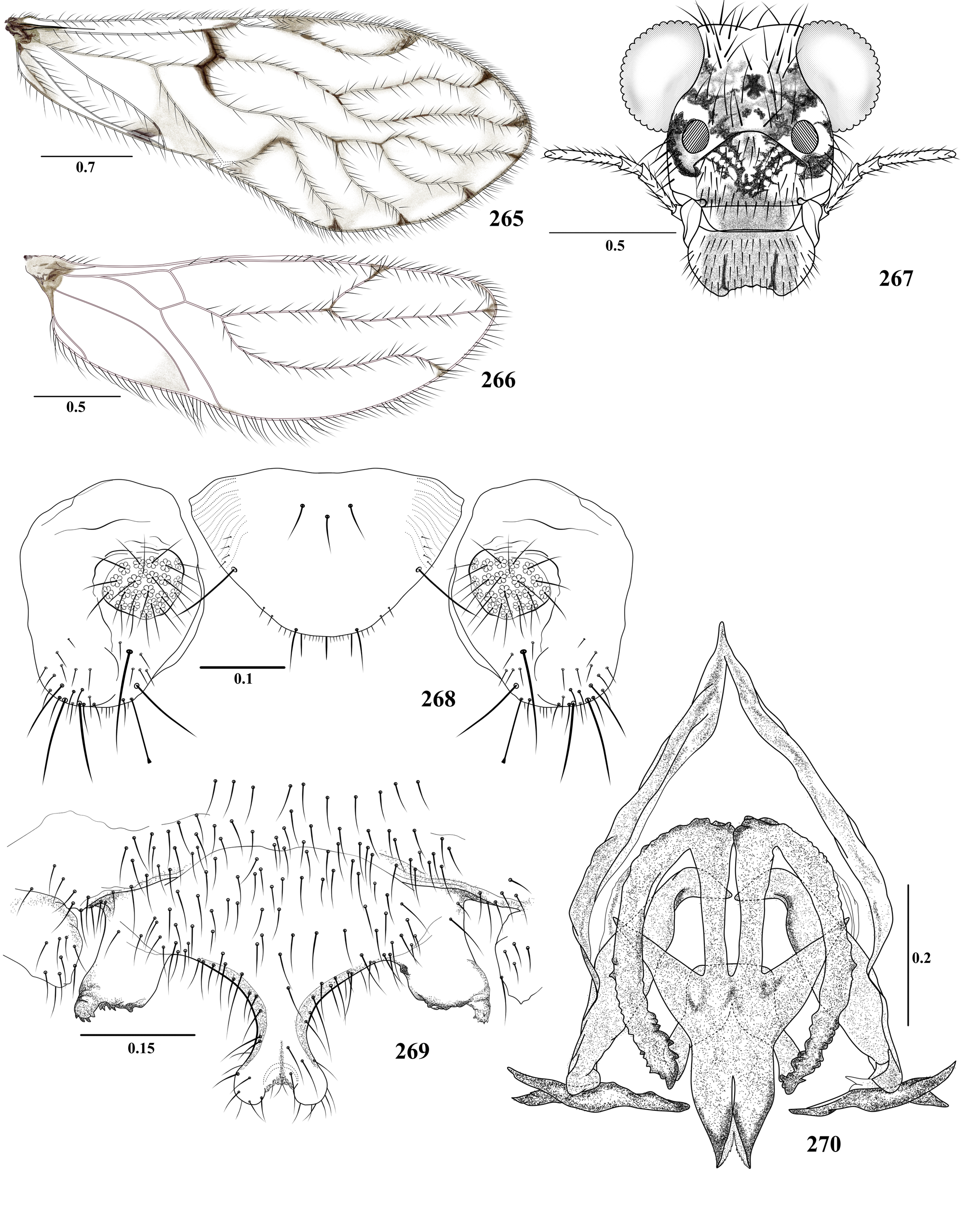
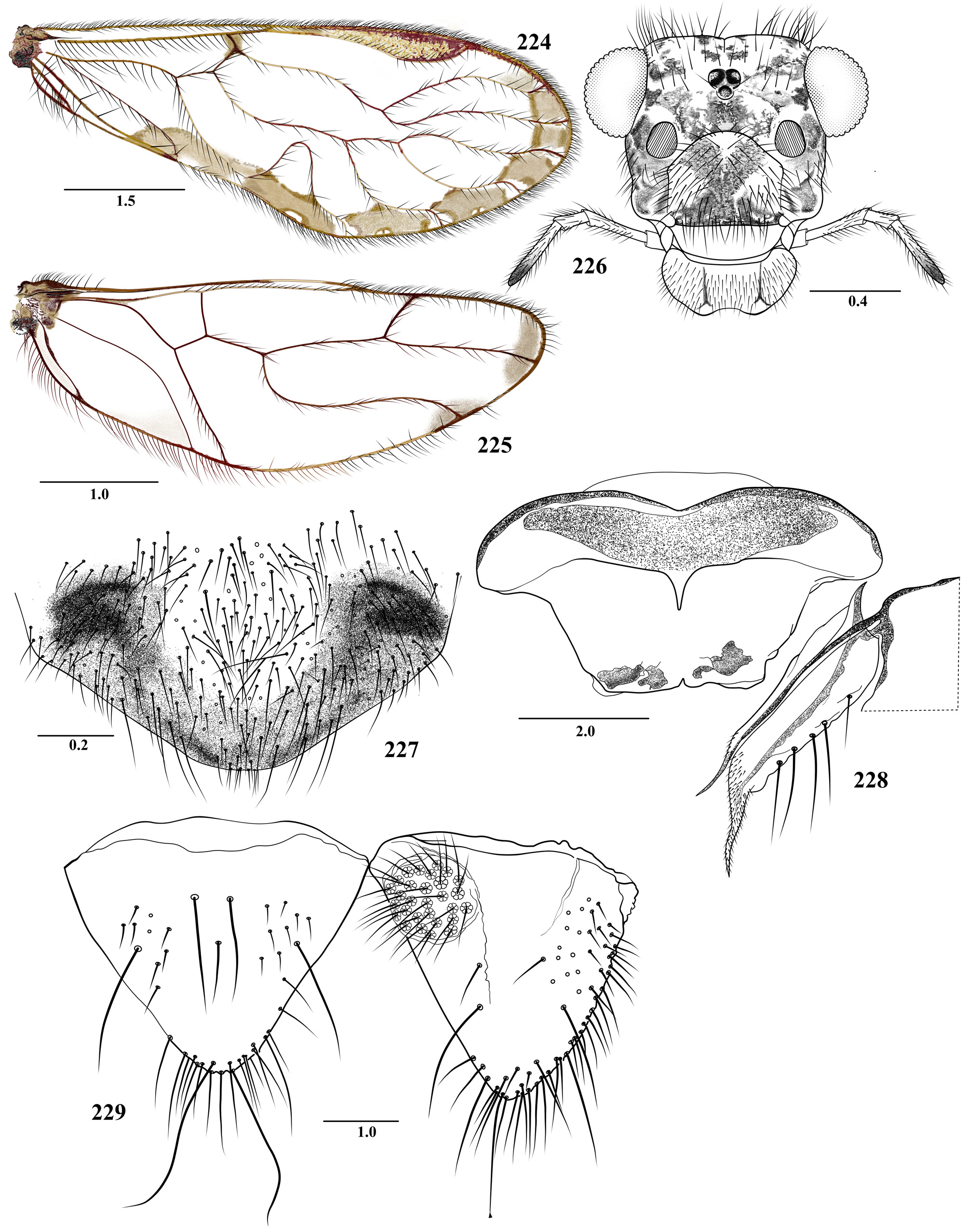
31. Central sclerite of hypandrium with a median, distally bilobed posterior projection, and a broad process on each postero lateral corner, each bearing a rounded lateral projection, with a row of short, sclerotized teeth along the posterior border ( Fig. 269 View FIGURES 265 – 270 ); mesal pair of endophallic sclerites with central, fused area arrow shaped, posteriorly bifid, anteriorly with two arms, each one straight in the middle, then bow-shaped, with outer border irregular distally bearing denticles along both sides, posterior endophallic sclerites transverse, slender, with inner ends pointed and outer ends bifid, acuminate ( Fig. 270 View FIGURES 265 – 270 )....................................................................................................... T. rugosa n. sp.
- Central sclerite of hypandrium anteriorly straight, with postero-lateral corners sclerotized, projected posteriorly; posterior border with two short projections in the middle, apically blunt, directed outwards, the space between the middle and the lateral projections denticulate, strongly sclerotized ( Fig. 73 View FIGURES 69 – 74 ); anterior pair of endophallic sclerites broadly M-shaped, mesal pair Xshaped, with posterior arms curved, acuminate; posterior pair close to the inner border of the external parameres, each half elongate, with a row of four teeth along the inner border ( Fig. 74 View FIGURES 69 – 74 )................................... T. bubuae n. sp.
32. Central sclerite of hypandrium with latero-caudal processes separated by a deep or shallow concavity ( Figs 13 View FIGURES 9 – 14 and 132 View FIGURES 128 – 133 ); posterior endophallic sclerites H-shaped ( Figs 14 View FIGURES 9 – 14 and 133 View FIGURES 128 – 133 )........................................................ 33
- Central sclerite of hypandrium with median processes, small or well developed ( Figs 31 View FIGURES 27 – 32 , 263 View FIGURES 259 – 264 ); mesal endophallic sclerites Y or H-shaped ( Figs 32 View FIGURES 27 – 32 and 264 View FIGURES 259 – 264 )............................................................................ 34
33. Central sclerite of hypandrium with two short, stout posterior projections, with a deep concavity between them ( Fig. 132 View FIGURES 128 – 133 ); two pairs of endophallic sclerites, anterior pair with inner arms slender, acuminate; outer arms club-shaped; mesal pair X-shaped, with posterior arms distally rounded, with an acuminate lateral projection ( Fig. 133 View FIGURES 128 – 133 ).................... T. embera n. sp.
- Central sclerite of hypandrium with two short, blunt ended posterior projections, with a narrow concavity between them ( Fig. 13 View FIGURES 9 – 14 ); three pairs of endophallic sclerites, mesal pair stout, H-shaped, anterior pair with inner arms straight, slender, acuminate, outer arms curved, stout ( Fig. 14 View FIGURES 9 – 14 )....................................................... T. anchicayaensis n. sp.
34. Forewing M1 with Y-shaped spur vein ( Fig. 27 View FIGURES 27 – 32 ); central sclerite of hypandrium oval, with a small, concave posterior projection in the middle ( Fig. 31 View FIGURES 27 – 32 ); anterior pair of endophallic sclerites with fan-shaped processes, postero-mesal pair dilated posteriorly, fork-shaped ( Fig. 32 View FIGURES 27 – 32 )...................................................................... T. arhuaca n. sp.
- Forewing M1 without spur vein ( Fig. 259 View FIGURES 259 – 264 ); central sclerite of hypandrium convex anteriorly, bilobed posteriorly, with apex widely rounded and closely spaced ( Fig. 263 View FIGURES 259 – 264 ); mesal endophallic sclerite transverse, H-shaped, with two long anterior arms, posterior arms straight, acuminate, and close together ( Fig. 264 View FIGURES 259 – 264 )................................. T. robustoides n. sp.
35. Forewing M3 simple.................................................................................. 36
- Forewing M3 branched, bi- or trifurcated ( Figs 305 View FIGURES 305 – 310 , 311 View FIGURES 311 – 316 , 317 View FIGURES 317 – 322 , 323 View FIGURES 323 – 328 )............................................. 60
36. Forewings with dark brown pigmentation proximally, sometimes with a wide, marginal pigmented band ( Figs 194 View FIGURES 194 – 199 and 200 View FIGURES 200 – 205 ); A2 reaching the wing margin; IX sternum convex, emarginate, with clearly defined antero-lateral lobe ( Figs 198 View FIGURES 194 – 199 and 205 View FIGURES 200 – 205 )...................................................................................................... 37
- Forewings hyaline, or slightly pigmented proximally, sometimes with wide marginal pigmented band; A2 reaching or not the wing margin; IX sternum variable........................................................................ 38
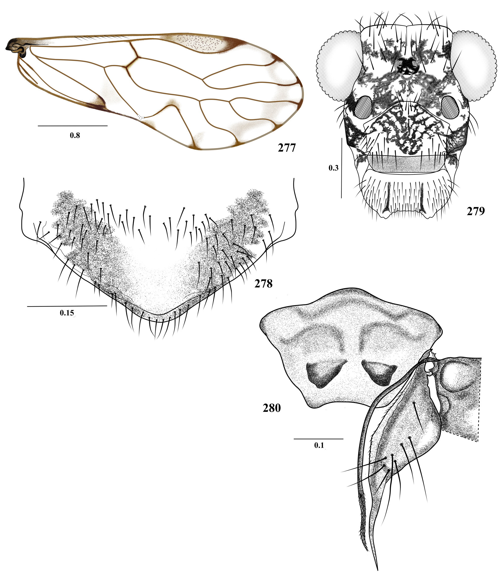
37. Forewings without marginal pigmented band, deeply pigmented proximally ( Fig. 200 View FIGURES 200 – 205 ); labral sclerites evident ( Fig. 202 View FIGURES 200 – 205 ); ninth sternum concave anteriorly in the middle ( Fig. 205 View FIGURES 200 – 205 ).......................................... T. lapayaensis n. sp.
- Forewing with broad marginal pigmented band from R4+5 to areola postica, with large dark brown proximal area, pterostigma with proximal and distal brown bands; A2 joining A1 ( Fig. 194 View FIGURES 194 – 199 ); labral sclerites not evident ( Fig. 196 View FIGURES 194 – 199 ); IX sternum trapeziform, peripherally pigmented ( Fig. 198 View FIGURES 194 – 199 )....................................................... T. korebaju n. sp.
38. Forewings with well defined pigmented marginal band, at least between R4+5 and Cu1b ( Figs 158 View FIGURES 158 – 163 , 224 View FIGURES 224 – 229 , and 281)........ 3 9
- Forewings hyaline or with pigmented marginal band not well defined, if it continues, then with hyaline or clear spaces near margin of the respective cells ( Figs 21 View FIGURES 21 – 26 , 33,122, 182, 242, and 277).............................................. 44
39. Forewings with pigmented marginal band without setae; areola postica with ratio length /width greater than 1.5, with strong concavity on the part distal to Cu1a ( Fig. 151 View FIGURES 146 – 151 ); IX sternum with two well defined areas, the anterior widened, with lateral lobes and leading edge convex, emarginate ( Fig. 162 View FIGURES 158 – 163 )............................................................. 40
- Forewings with pigmented marginal band with setae; areola postica with ratio length/width smaller than 1.5; IX sternum variable............................................................................................... 42
40. Forewing with A2 reaching the wing margin ( Figs 224 View FIGURES 224 – 229 and 281 View FIGURES 281 – 286 ); IX sternum with anterior area distinctly delimited ( Figs 228 View FIGURES 224 – 229 and 286 View FIGURES 281 – 286 ), with oval laminar structure in central area........................................................ 41
- Forewing with A2 not reaching the wing margin ( Fig. 158 View FIGURES 158 – 163 ); IX sternum slender posteriorly, with anterior, transverse, pigmented narrow band, without central laminar structure ( Fig. 162 View FIGURES 158 – 163 )..................................... T. guane n. sp.
41. Ninth sternum with three well delimited areas, a pigmented transverse anterior one, a mesal area projected posteriorly in the middle, with one transverse sickle-shaped pigmented area on each side of the longitudinal midline, and one mostly hyaline posterior area ( Fig. 286 View FIGURES 281 – 286 )................................................................. T. sevillaensis n. sp.
- IX sternum broader posteriorly, with the anterior, transverse pigmented band wider ( Fig. 228 View FIGURES 224 – 229 ), posterior area distinctly wide, trapezoida.............................................................................. T. matildae n. sp.
42. Forewings with transverse pale brown band from rs-m to apex of cell cup; veins M1 to Cu1b ending in a hyaline fenestra, with dark sides ( Fig. 212 View FIGURES 212 – 217 ); IX sternum with a well-defined, anterior transverse area and two posterior lobes, anterior margin distinctly emarginated ( Fig. 217 View FIGURES 212 – 217 ).............................................................. T. lithophila n. sp.
- Forewings without transverse pale brown band from rs-m to apex of cell cup, longitudinal veins with brown areolae at setal insertions ( Figs 81 View FIGURES 81 – 86 and 140 View FIGURES 140 – 145 ); IX sternum variable ( Figs 86 View FIGURES 81 – 86 and 145 View FIGURES 140 – 145 )............................................. 43
43. IX sternum with three well defined areas, the anterior one oval; mesal area with an anterior V-shaped concavity, and a posterior, trapeziform pigmented process; posterior area membranous, unpigmented ( Fig. 86 View FIGURES 81 – 86 )................. T. calima n. sp.
- IX sternum trapeziform, with a distinct, pigmented transverse band, projected posteriorly in the middle ( Fig. 145 View FIGURES 140 – 145 )............................................................................................... T. felidiaensis n. sp.
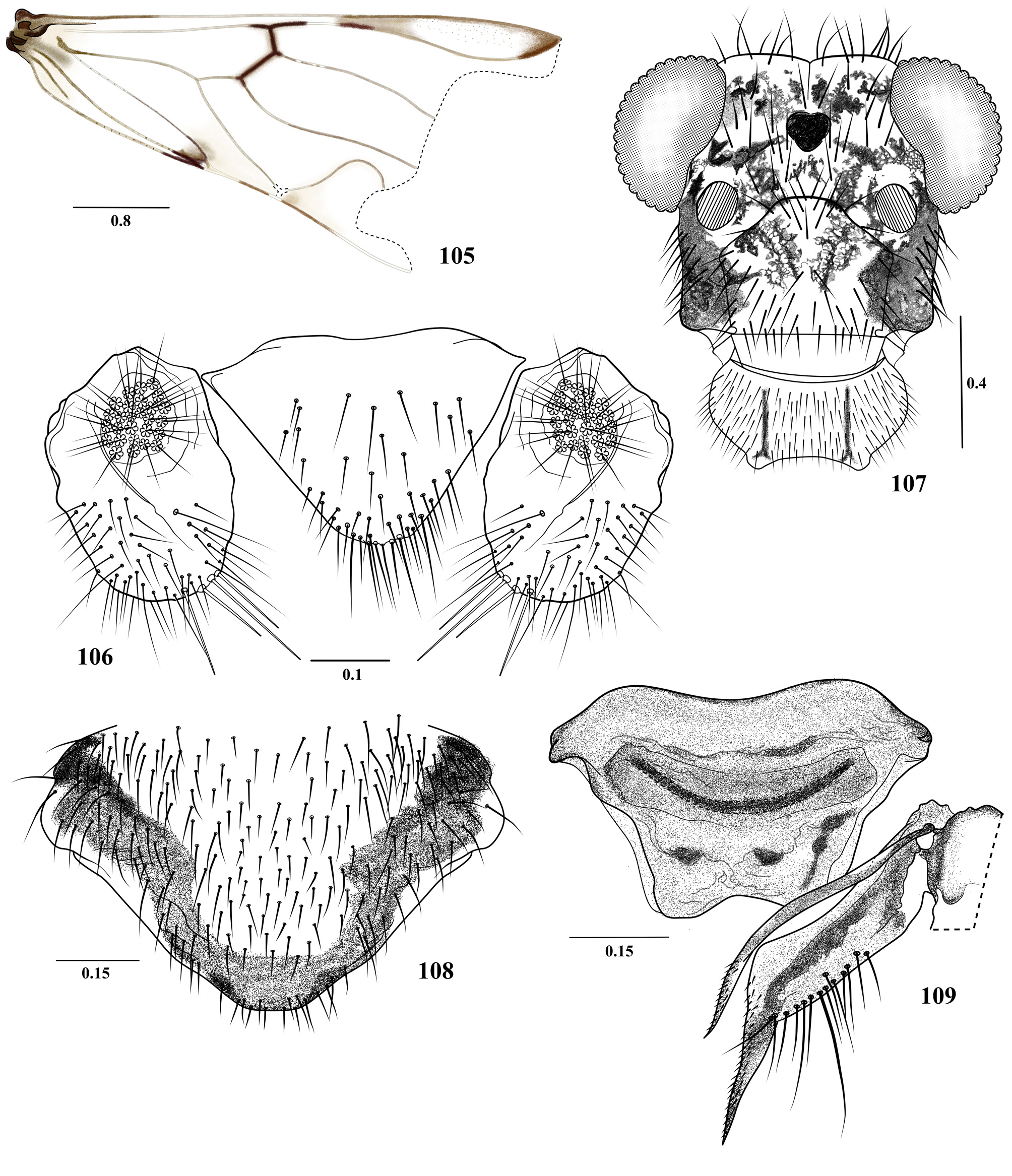
44. Forewings with vein A2 not reaching the wing margin....................................................... 45
- Forewings with vein A2 reaching the wing margin.......................................................... 57
45. Forewings hyaline, membrane with spots at the ends of the veins, with or without short spur veins on margin, on cells m2 and m3 ( Figs 21 View FIGURES 21 – 26 and 122 View FIGURES 122 – 127 ); IX sternum with widened anterior area, rounded edges and front side emarginated ( Figs 26 View FIGURES 21 – 26 and 127 View FIGURES 122 – 127 )..................................................................................................... 46
- Forewings hyaline or with pigmented areas, without spur veins on margin ( Figs 33 View FIGURES 33 – 38 , 182 View FIGURES 182 – 187 , 242 View FIGURES 242 – 246 , and 277); IX sternum with anterior area variable..................................................................................... 47
46. Forewings with short spur veins on margin, on cells m2 and m3 ( Fig. 21 View FIGURES 21 – 26 ); IX sternum without sclerotized or pigmented area in anterior area ( Fig. 26 View FIGURES 21 – 26 )..................................................................... T. andaqui n. sp.
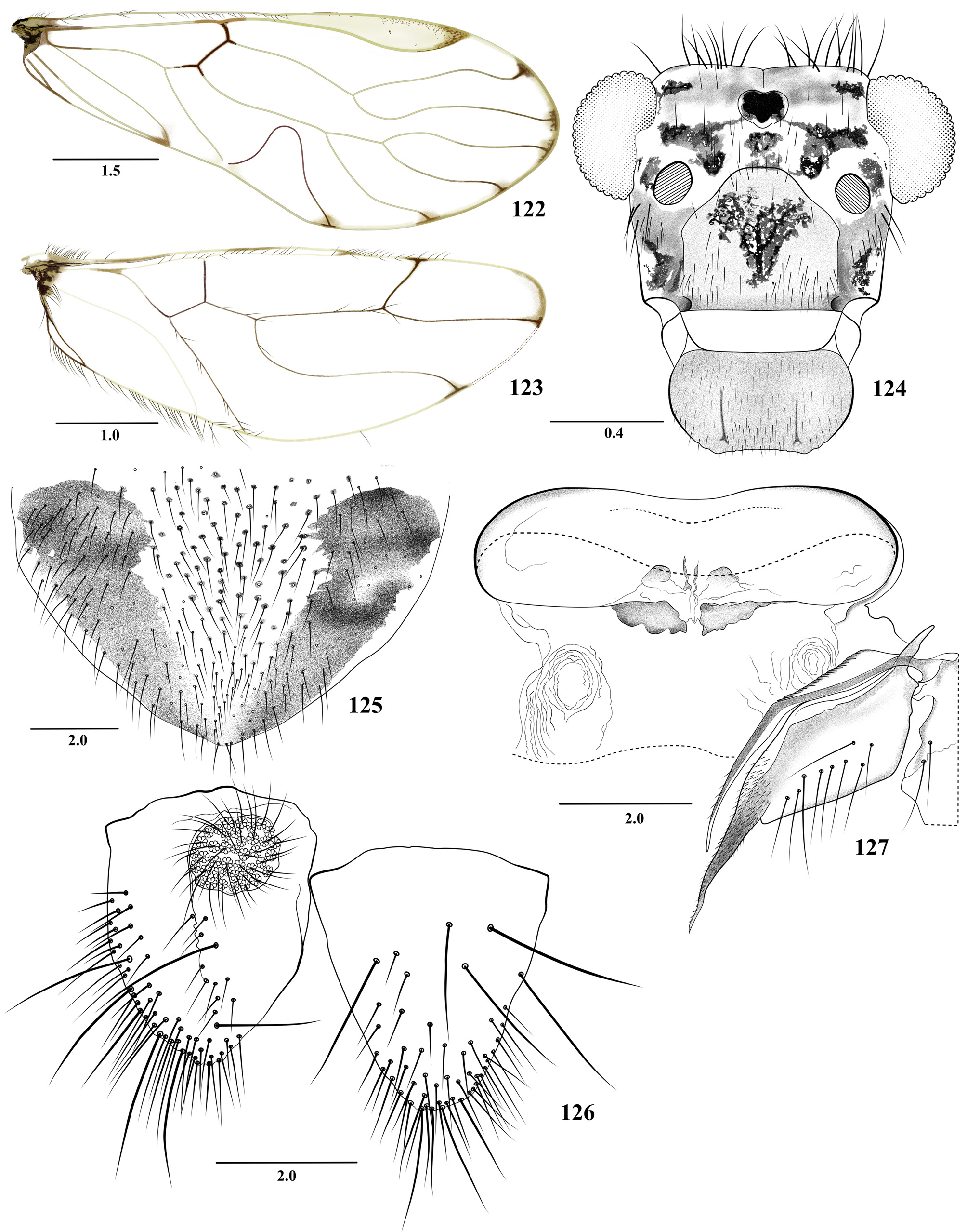
- Forewings without spur veins as above ( Fig. 122 View FIGURES 122 – 127 ); IX sternum with two sclerotized areas in the posterior margin of the anterior area ( Fig. 127 View FIGURES 122 – 127 )............................................................................ T. dimitrii n. sp.
47. Forewings with a transverse dark or pale brown band from rs-m to apex of cell cup ( Figs 63 View FIGURES 63 – 68 and 370 View FIGURES 370 – 373 ); distal part of Cu1a with or without distinct emargination......................................................................... 48
- Forewings without a transverse dark brown band between rs-m to apex of the cell cup ( Figs 182 View FIGURES 182 – 187 , 277 View FIGURES 277 – 280 ); distal part of areola postica almost straight, sometimes emarginated................................................................ 49 48. IX sternum broadly pentagonal ( Fig. 68 View FIGURES 63 – 68 ); Cula concave in the middle ( Fig. 63 View FIGURES 63 – 68 )....................... T. bicornuta n. sp.
- IX sternum oval ( Fig. 373 View FIGURES 370 – 373 ); Cu1a almost straight ( Fig. 370 View FIGURES 370 – 373 ).............................. T. colombiana García Aldrete
49. IX sternum trapeziform, with convex anterior margin ( Figs 186 View FIGURES 182 – 187 and 280 View FIGURES 277 – 280 )........................................ 50
- IX sternum cup-shaped, with anterior margin convex or straight ( Figs 55 View FIGURES 51 – 56 , 103 View FIGURES 99 – 104 , 109 View FIGURES 105 – 109 , 157 View FIGURES 152 – 157 , 175 View FIGURES 170 – 175 , 298 View FIGURES 293 – 298 and 303 View FIGURES 299 – 304 ); if trapeziform, then anterior margin fully emarginate..................................................................... 51
50. IX sternum broadly trapeziform, convex anteriorly; posteriorly with a triangular, sclerotized area on each side of longitudinal midline ( Fig. 280 View FIGURES 277 – 280 ); forewings hyaline, with a submarginal pale brown band from R2+3 to M3; Cu1a straight ( Fig. 277 View FIGURES 277 – 280 )............................................................................................. T. sarmaca n. sp.
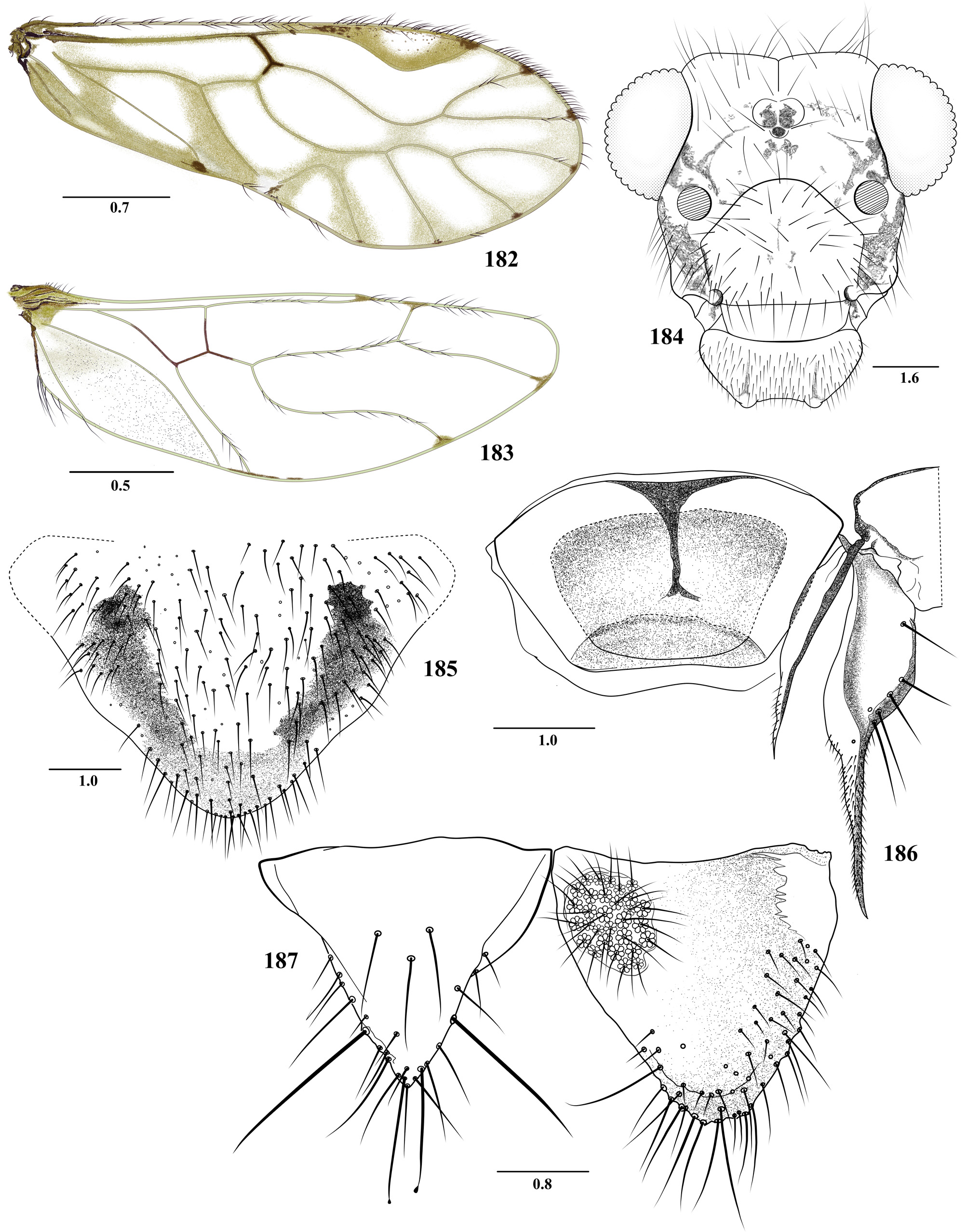
- IX sternum trapeziform, with pigmented band proximally in the middle ( Fig. 186 View FIGURES 182 – 187 ); forewings hyaline, with brown pigmented areas as in figure 182......................................................................... T. inga n. sp.
51. IX sternum quadrangular, with oval anterior area and cup-shaped posterior area ( Fig. 103 View FIGURES 99 – 104 ); Cu1a concave in the middle ( Fig. 99 View FIGURES 99 – 104 )..................................................................................... T. cantatis n. sp.
- IX sternum subquadrangular, without oval anterior area ( Figs 55 View FIGURES 51 – 56 , 109 View FIGURES 105 – 109 , 157 View FIGURES 152 – 157 , 298 View FIGURES 293 – 298 , and 303); Cu1a almost straight or gently concave ( Figs 45 View FIGURES 45 – 50 , 152 View FIGURES 152 – 157 , 164 View FIGURES 164 – 169 , 293 View FIGURES 293 – 298 , and 299).................................................................... 52
52. IX sternum quadrangular: anterior area projected posteriorly in the middle ( Fig. 298 View FIGURES 293 – 298 ); subgenital plate with well defined pigmented area ( Fig. 297 View FIGURES 293 – 298 )................................................................... T. yanacona n. sp.
- IX sternum not as above; subgenital plate with pigmented area variable.......................................... 53
53. IX sternum cup-shaped, pigmented, anterior margin slightly convex, almost straight, with ventro-anterior V-shaped pigmented area ( Fig. 55 View FIGURES 51 – 56 ), posteriorly rounded, with two rounded processes........................................ T. awa n. sp.
- IX sternum cup or trapeziform-shaped, without sclerotized areas as above........................................ 54
54. IX sternum cup-shaped, anteriorly wide; anterior and posterior margins almost straight, with two sclerotized large sublateral areas distally ( Fig. 157 View FIGURES 152 – 157 ).............................................................. T. garciamarquezi n. sp.
- IX sternum cup-shaped or trapeziform, with anterior margin distinctly emarginate, or without sclerotized areas as above... 55
55. IX sternum trapeziform, wider anteriorly, slightly concave in the middle, narrower posteriorly, deeply concave in the middle ( Fig. 109 View FIGURES 105 – 109 ); a mesal, transverse, bow-shaped pigmented band, and one small pigmented area posteriorly, on each side of the longitudinal midline........................................................................... T. caribe n. sp.
- IX sternum trapeziform, anterior margin clearly emarginated ( Figs 175 View FIGURES 170 – 175 and 303 View FIGURES 299 – 304 )................................... 56
56. IX sternum cup-shaped, concave anteriorly, projected posteriorly, with membranous areas on sides ( Fig. 175 View FIGURES 170 – 175 )..................................................................................................... T. huitota n. sp.
- IX sternum trapeziform, anterior margin widely emarginated; posteriorly without lateral concavity or oval areas as in T. huitota ( Fig. 303 View FIGURES 299 – 304 )........................................................................ T. erwini Silva-Neto et al.
57. Forewing M1 with a Y-shaped spur vein ( Fig. 33 View FIGURES 33 – 38 ); IX sternum broadly trapeziform, with a transverse, pigmented band anteriorly, and an irregular pigmented area on each postero-lateral corner ( Fig. 38 View FIGURES 33 – 38 )......................... T. arhuaca n. sp.
- Forewing M1 without spur vein ( Figs 236 View FIGURES 236 – 241 , 242 View FIGURES 242 – 246 and 271 View FIGURES 271 – 276 ); IX sternum variable ( Figs 241 View FIGURES 236 – 241 , 246 View FIGURES 242 – 246 , and 276)................ 58
58. Forewings with slender, pale brown marginal band; areola postica slanted posteriorly ( Fig. 242 View FIGURES 242 – 246 ); IX sternum wider anteriorly, concave on the sides, and anteriorly and posteriorly; a transverse pigmented band along anterior border ( Fig. 246 View FIGURES 242 – 246 )................................................................................................. T. motilona n. sp.
- Forewings not as above, areola postica not slanted ( Figs. 236 View FIGURES 236 – 241 and 271 View FIGURES 271 – 276 ); IX sternum variable ( Figs 241 View FIGURES 236 – 241 and 276 View FIGURES 271 – 276 ).......... 59
59. IX sternum long, trapeziform, wider anteriorly, slightly concave in the middle ( Fig. 276 View FIGURES 271 – 276 )................. T. rugosa n. sp.
- IX sternum cup-shaped, anterior margin slightly emarginated ( Fig. 241 View FIGURES 236 – 241 )........................... T. mocoaensis n. sp.
60. Forewings hyaline, without dark marginal band ( Figs 335 View FIGURES 335 – 340 , 353 View FIGURES 353 – 358 and 359 View FIGURES 359 – 364 ); IX sternum of two large areas, anterior one with lateral margins oval, anterior margin emarginate centrally, with mesal, narrow and elongate concavity projected mesally; epiproct with oval bulge in mesal area, sometimes not evident in dorsal view ( Figs 316 View FIGURES 311 – 316 , 358 View FIGURES 353 – 358 and 364 View FIGURES 359 – 364 )......................... 61
-..... Forewings with pale brown area proximally, with dark marginal band from R2+3 to Cu1a ( Figs 317 View FIGURES 317 – 322 , 329 View FIGURES 329 – 334 , 341 View FIGURES 341 – 346 and 347 View FIGURES 347 – 352 ); pterostigma brown, with cross pale brown spot from rs-m to anal margin; IX sternum not as above; epiproct without oval bulge in mesal area ( Figs 321 View FIGURES 317 – 322 , 333 View FIGURES 329 – 334 , 344 View FIGURES 341 – 346 and 350 View FIGURES 347 – 352 )................................................................ 64
61. IX sternum posteriorly without longitudinal grooves or clearly delimited by lateral sclerotized area, posterior border emarginate ( Figs 364 View FIGURES 359 – 364 and 373 View FIGURES 370 – 373 )................................................................................. 62
- IX sternum posteriorly with longitudinal grooves, posterior border rounded, not emarginate ( Figs 316 View FIGURES 311 – 316 and 358 View FIGURES 353 – 358 ).......... 63
62. IX sternum with posterior area pigmented, thicker than the anterior one, with a strongly sclerotized latero-posterior band, and a small, strongly pigmented oval area mesally on each side ( Fig. 373 View FIGURES 370 – 373 )..................... T. lamasoides Silva-Neto et al.
- IX sternum with posterior area not pigmented, without a strongly sclerotized latero-posterior band, slightly pigmented or without pigmented oval area mesally on each side ( Fig. 364 View FIGURES 359 – 364 )................................... T. lamasi Silva-Neto et al.
63. IX sternum posteriorly with slightly developed longitudinal grooves mesally ( Fig. 358 View FIGURES 353 – 358 ), V2 +3 proximally wide, V3 with widely rounded apex........................................................................ T. furcata New
- IX sternum posteriorly with many longitudinal grooves, elongated mesally, more than in T. furcata ( Fig. 316 View FIGURES 311 – 316 ); v2 +3 proximally not as wide as above, v2 with apex less rounded.............................................. T. furcatoides n. sp.
64. A2 not reaching the wing margin, IX sternum with elongate lateral and sclerotized brown area, lateral borders well sclerotized ( Figs 322 View FIGURES 317 – 322 and 334 View FIGURES 329 – 334 ).................................................................................... 65
- A2 reaching the wing margin; IX sternum without sclerotized lateral area, lateral borders well sclerotized, irregularly oval or cup-shaped ( Figs 346 View FIGURES 341 – 346 and 352 View FIGURES 347 – 352 ).......................................................................... 66
65. IX sternum with semi-oval anterior area, delimited by latero-posterior brown slender bands; posteriorly emarginate ( Fig. 322 View FIGURES 317 – 322 ); fore- coxa pale brown, with brown ochre spot, middle and posterior coxae cream..................... T. huilaensis n. sp.
- IX sternum with trapeziform; anterior area delimited by latero-posterior brown bands; anterior border emarginate ( Fig. 334 View FIGURES 329 – 334 ); fore- and mid coxae dark brown, hind- coxae cream....................................... T. lamensuraensis n. sp.
66. IX sternum shield-shaped, wider anteriorly, with lateral borders limited by a sclerotized band, anterior border gently emarginated; latero-posteriorly with two oval, projected membranous areas ( Fig. 346 View FIGURES 341 – 346 )................ T. otunquimbayaensis n. sp.
- IX sternum broadly oval, lateral borders limited by a sclerotized band, anterior border projected in the middle, posterior border with corners protruding ( Fig. 352 View FIGURES 347 – 352 )............................................................ T. sarriae n. sp.


No known copyright restrictions apply. See Agosti, D., Egloff, W., 2009. Taxonomic information exchange and copyright: the Plazi approach. BMC Research Notes 2009, 2:53 for further explanation.
|
Kingdom |
|
|
Phylum |
|
|
Class |
|
|
Order |
|
|
Family |