
Tuberculobasis
|
publication ID |
https://doi.org/10.5281/zenodo.187806 |
|
DOI |
https://doi.org/10.5281/zenodo.4391361 |
|
persistent identifier |
https://treatment.plazi.org/id/1312774B-FFD7-FFE5-FF04-FE98FA3CF906 |
|
treatment provided by |
Plazi |
|
scientific name |
Tuberculobasis |
| status |
|
Key to males of Tuberculobasis View in CoL
(Male of T. cardinalis unknown)
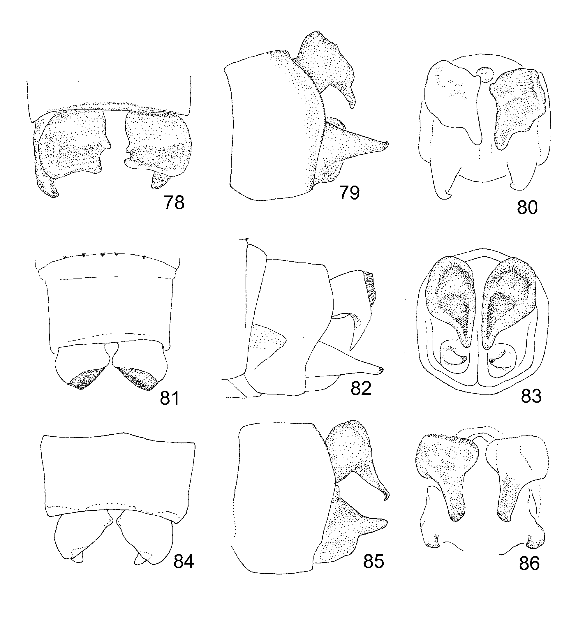
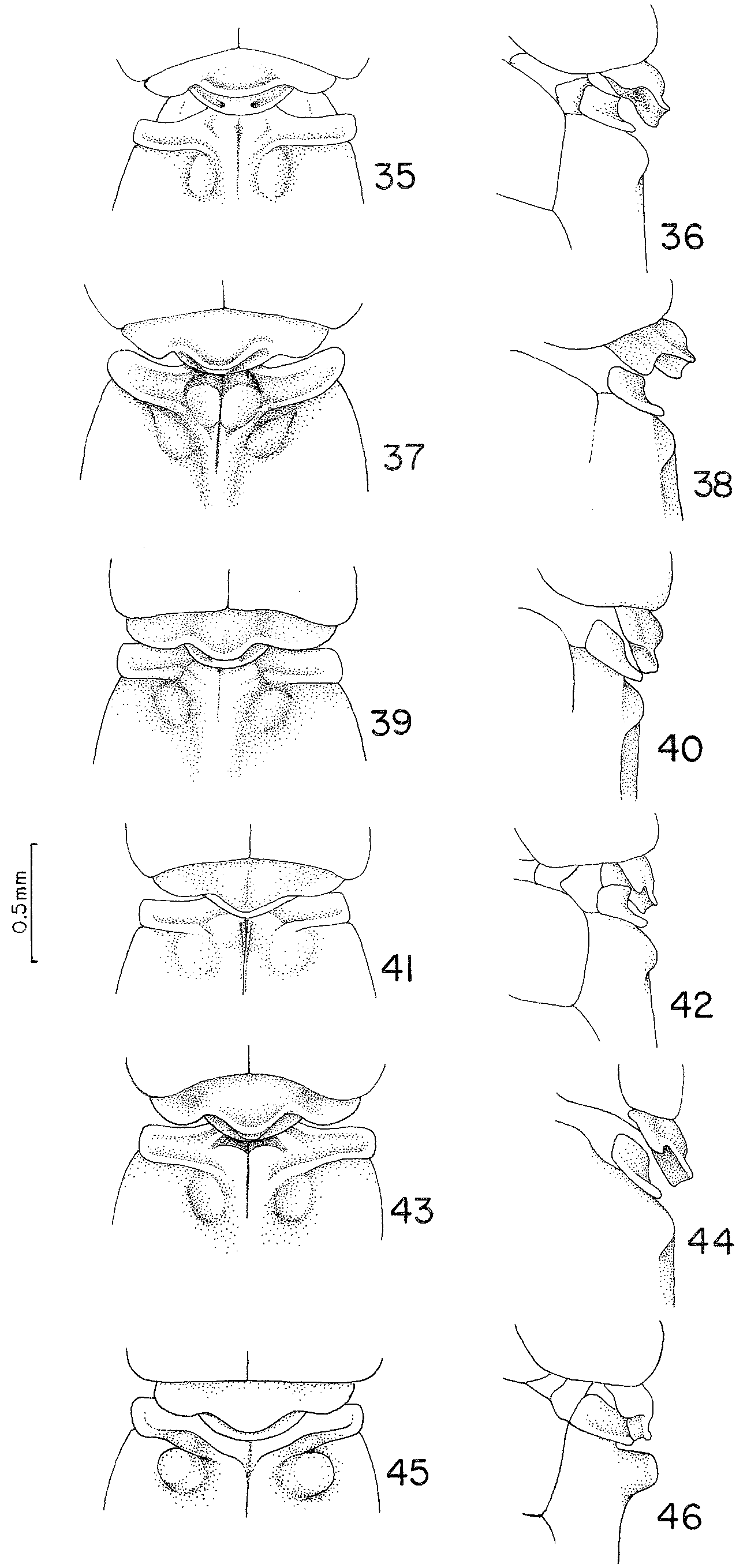
1. Paraproct subequal or slightly longer than cercus ( Figs 79, 82, 85 View FIGURES 78 – 86 ). Mesepisternal tubercles mamiliform and low (<0.82 mm) ( Figs 36, 38, 40, 42, 44 View FIGURES 35 – 46 )............................................................................................................................ 2
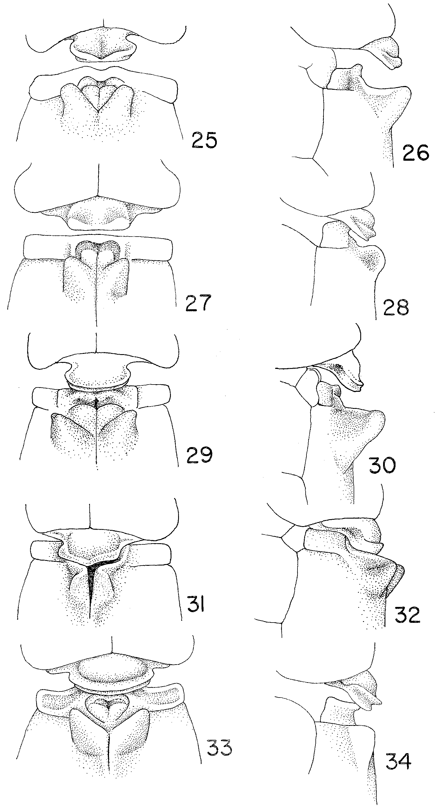
1´. Paraproct much longer than cercus ( Figs 57–58, 60–61, 64 View FIGURES 57 – 65 , 67). Mesepisternal tubercles not mammiliform and high (>1.41 mm: Figs 26, 28, 30, 32, 34 View FIGURES 25 – 34 , 46 View FIGURES 35 – 46 ) ....................................................................................................................... 6
2. Mesepisternal tubercle in dorsal view tear-shaped, tapering to a fine tip antero-distally ( Fig. 37 View FIGURES 35 – 46 ). Mesostigmal plates curved and directed anteriorly ( Fig. 37 View FIGURES 35 – 46 ) Rondônia State, Brazil................................................................. T. karitiana View in CoL
2´. Mesepisternal tubercles not tear-shaped. Mesostigmal plates straight ( Figs. 35, 39, 41, 43, 45 View FIGURES 35 – 46 ) ............................... 3
3. Hind part of dorsum of S10 elevated ( Fig. 77 View FIGURES 69 – 77 ). Median lobe of hind prothoracic lobe as broad as lateral lobes taken together ( Fig. 35 View FIGURES 35 – 46 ). Mesepisternum with metallic luster Suriname................................................................ T. geijskesi View in CoL
3´. Hind part of dorsum of S10 not elevated ( Fig. 80, 83, 86 View FIGURES 78 – 86 ). Median lobe of hind prothoracic lobe narrower than lateral lobes taken together ( Figs 41, 43 View FIGURES 35 – 46 ). Mesepisternum lacking metallic luster........................................................... 4
4. Medial margin of cercus in posterior view with a distinct dorsal lobe making the ventral part of medial margin appear markedly concave ( Fig. 86 View FIGURES 78 – 86 ) Venezuela, Brazil............................................................................... T. yanomami View in CoL
4´. Medial margin of cercus in posterior view almost straight with no dorsal lobe ( Fig. 83 View FIGURES 78 – 86 ) or with only a small one ( Fig. 80 View FIGURES 78 – 86 ), with ventral part of medial margin slightly convex to slightly concave................................................................ 5
5. Ventral process of cercus in lateral view a triangular plate tapering ventrally into a fine straight tip ( Fig. 82 View FIGURES 78 – 86 ). Penis ( Fig. 92 View FIGURES 87 – 94 ) with proximal lateral lobe digitiform. Pará State, Brazil...................................................................... T. tirio View in CoL
5´. Ventral process of cercus in lateral view digitiform with tip blunt and slightly curved anteriorly ( Fig. 79 View FIGURES 78 – 86 ). Penis ( Fig. 91 View FIGURES 87 – 94 ) with proximal lateral lobe triangular. Brazil, Venezuela................................................................... T. mammilaris View in CoL
6. Mesepisternal tubercles with their bases well separated from mid-dorsal carina; lateral lobes of hind prothoracic lobe well developed ( Fig. 45 View FIGURES 35 – 46 ). Roraima State, Brazil...................................................................................... T. macuxi View in CoL
6 ´. Mesepisternal tubercles with their bases adjacent to mid-dorsal carina; lateral lobes of hind prothoracic lobe poorly developed or absent ( Figs 25, 27, 29, 31, 33 View FIGURES 25 – 34 ) ............................................................................................................... 7
7. Mesepisternal tubercles subparallel ( Figs 31, 32 View FIGURES 25 – 34 ) with the anterior border connected with mesostigmal plate by a vertical curved plate ( Fig. 31 View FIGURES 25 – 34 ). Amazonas, Pará, and Rondônia States, Brazil.............................................. T. inversa View in CoL
7´. Mesepisternal tubercles strongly divergent ( Figs 25, 27, 29, 33 View FIGURES 25 – 34 ) with anterior border not connected with mesostigmal plate by a vertical curved plate ( Figs 25–30, 33, 34 View FIGURES 25 – 34 ) ............................................................................................ 8
8. Cercus in posterior view ( Fig. 74 View FIGURES 69 – 77 ) with a dorsolateral semilunar cavity limited by a denticulated border. Colombia and Venezuela.............................................................................................................................................................. 8.
8`. Cercus in posterior view ( Figs. 59, 62, 65 View FIGURES 57 – 65 ) with no dorsolateral semilunar cavity ...................................................... 9
9. Ventral process of cercus in posterior view digitiform with medial margin curved ( Fig. 62 View FIGURES 57 – 65 ). SE Brazil. T. costalimai View in CoL
9´. Ventral process of cercus in posterior view subtriangular with medial margin straight ( Figs. 59, 65 View FIGURES 57 – 65 ) ...................... 10
10. Mesepisternal tubercles, high (2.36 mm), with the apex subtruncated ( Fig. 25 View FIGURES 25 – 34 ). Dorsal lip of medin lobe of hind prothoracic lobe slightly elevated, medially fused with ventral lip ( Fig. 25 View FIGURES 25 – 34 ). Anterior margin of mesostigmal plate with a median depression ( Fig. 25 View FIGURES 25 – 34 ). Rondônia State, Brazil...................................................................................... T. arara View in CoL
10´. Mesepisternal tubercles very high (2.83 mm), conical with the apex not subtruncated, directed laterally ( Fig. 29 View FIGURES 25 – 34 ). Dorsal lip of hind prothoracic lobe not elevated nor fused with ventral tip ( Fig. 29 View FIGURES 25 – 34 ). Anterior margin of mesostigmal plate with no median depression ( Fig. 29 View FIGURES 25 – 34 ). São Paulo State, Brazil.............................................................. T. guarani View in CoL


No known copyright restrictions apply. See Agosti, D., Egloff, W., 2009. Taxonomic information exchange and copyright: the Plazi approach. BMC Research Notes 2009, 2:53 for further explanation.
|
Kingdom |
|
|
Phylum |
|
|
Class |
|
|
Order |
|
|
Family |
|
|
Genus |