
Brachymeria trinidadensis (Narendran & Varghese)
|
publication ID |
https://doi.org/10.11646/zootaxa.5092.4.2 |
|
publication LSID |
lsid:zoobank.org:pub:D95A8BD9-8BA5-4825-BEA8-57556C80CBAD |
|
DOI |
https://doi.org/10.5281/zenodo.5915181 |
|
persistent identifier |
https://treatment.plazi.org/id/2F0D87A7-CA1B-FFC8-FF5A-FC1CFF5CE3E8 |
|
treatment provided by |
Plazi |
|
scientific name |
Brachymeria trinidadensis (Narendran & Varghese) |
| status |
|
Brachymeria trinidadensis (Narendran & Varghese) View in CoL
Thaumatelia trinidadensis Narendran & Varghese, 1989: 46‒47 View in CoL , figs 12‒18; combination by Halstead, 1991: 952.
Type material. Holotype, ♀ (photographs examined, Figs 1–4 View FIGURES 1–5 )—“ Holotype; Trinidad, Date ?, WI; Aug. Busck Collector; ♀ Thaumetelia trinidadensis SP. N, DET., NARENDRAN & THRESIAMA (VARGHESE), 1988; USNMENT 01559231” ( Fig. 5 View FIGURES 1–5 ).
Narendran & Varghese (1989) listed the type locality as “ Trinidad ( Argentina)”, but the type labels indicate otherwise .
Condition of holotype. Pinned. Right wings absent, left scape (lost after initial imaging, Miles Zhang, pers. comm.).
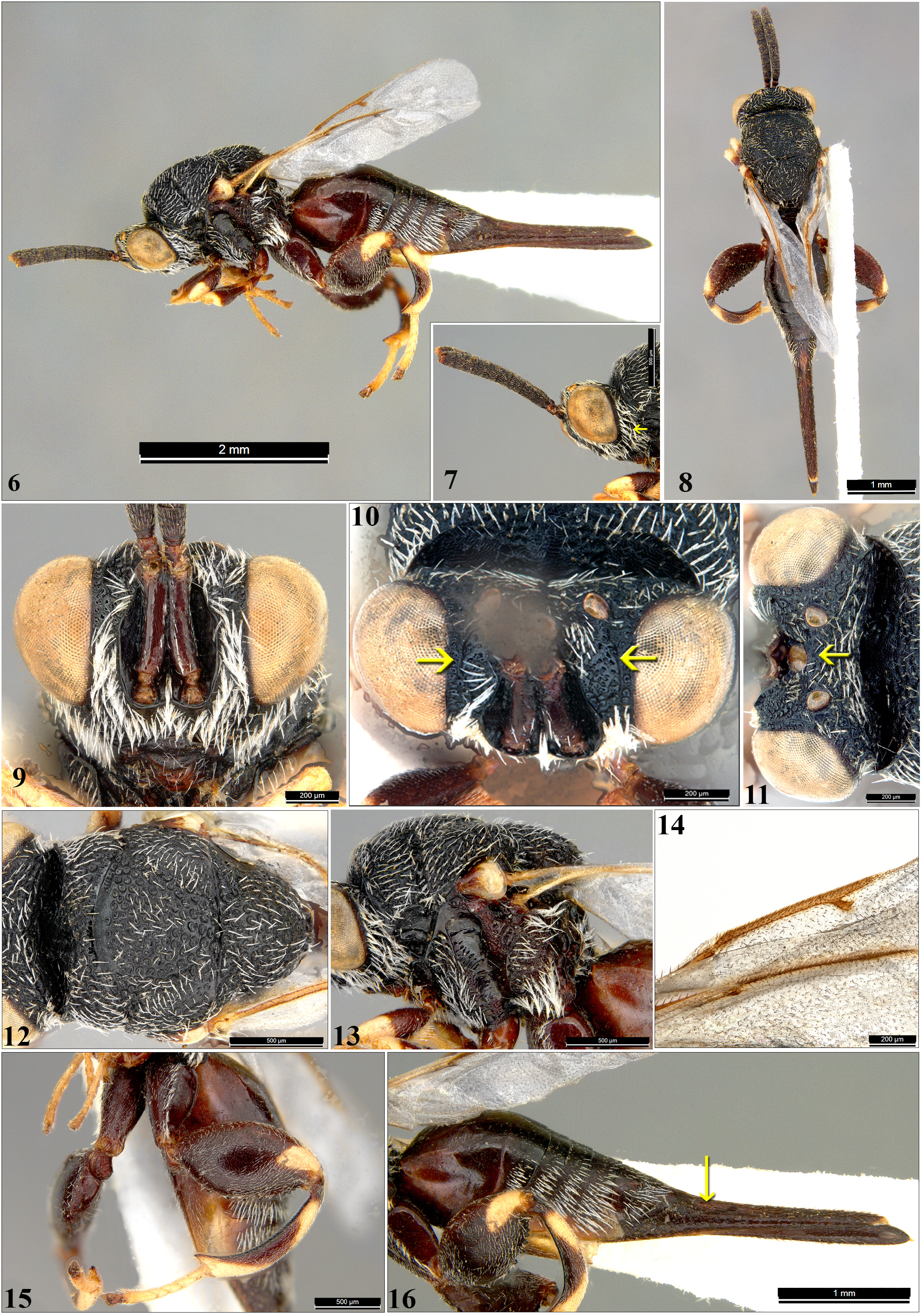
Other specimen examined. ♀, mounted on triangular card ( Figs 6–16 View FIGURES 6–16 )— India: Kerala, Kozhikode district, Elathur ( 11°19’32.5”N 75°44’30.8”E, 23.0 m above mean sea level), collected near subterranean tunnel of Bembix sp. , 25.vii.2020, coll. C. Bijoy. GoogleMaps
Diagnosis. Among known described species, females of B. trinidadensis are uniquely characterized by a conspicuously long syntergum, comprising 0.4× the length of the metasoma ( Figs 1, 4 View FIGURES 1–5 , 6, 8, 16 View FIGURES 6–16 ) and being about 4.6× as long as its basal width in dorsal view ( Figs 4 View FIGURES 1–5 , 8 View FIGURES 6–16 ) or basal height in lateral view ( Figs 1 View FIGURES 1–5 , 6, 16 View FIGURES 6–16 ) in combination with the head lacking a transverse carina on the vertex posterior to the median ocellus ( Figs 3, 4 View FIGURES 1–5 , 10, 11 View FIGURES 6–16 ). Other diagnostic features include: metasoma more than 1.8× as long as combined length of head and mesosoma ( Figs 1, 4 View FIGURES 1–5 , 6, 8 View FIGURES 6–16 ); head with post-orbital carina indicated, not reaching genotemporal margin (hardly visible beneath dense pilosity); hind femur reddish brown with apical yellow patch ( Figs 1 View FIGURES 1–5 , 6, 15, 16 View FIGURES 6–16 ); hind tibia subbasally and apically yellow with remainder reddish brown ( Figs 1 View FIGURES 1–5 , 6, 15 View FIGURES 6–16 ).
Redescription based on Indian female ( Figs 6–16 View FIGURES 6–16 ). Body length 5.52 mm; length of fore wing 2.79 mm.
Colour. Body black except as follows: eye reflective yellow; ocelli yellow; scape and pedicel reddish-brown; pleura dark brown; tegula yellow with base reddish-brown; coxae reddish-brown (hind coxa paler); fore and mid femora reddish-brown with yellow apical patch, hind femur reddish-brown with dorsoapical yellow patch; fore and mid tibiae yellow with median reddish-brown patch, hind tibia reddish-brown with subbasal and apical yellow patch; tarsi pale yellow; metasoma reddish-brown, but terga paler ventrally.
Head. Head in frontal view ( Fig. 9 View FIGURES 6–16 ) 1.5× as long as wide; densely and coarsely rugose-punctate, sculpture hidden beneath dense white setae on lower face, the frons and vertex less setose; scrobal margin conspicuously produced, distinctly carinate; scrobe deep, surface rugose-reticulate; frons with strong, uneven carina between each eye and scrobe, but the two carinae not curved and continued mesad dorsally behind median ocellus ( Figs 10, 11 View FIGURES 6–16 ); side of face without preorbital carina; post-orbital carina indicated, not reaching genotemporal margin ( Fig. 7 View FIGURES 6–16 ); eye 3.1× as long as malar space in profile ( Fig. 7 View FIGURES 6–16 ); head in dorsal view 1.1× as wide as mesosoma ( Figs 11, 12 View FIGURES 6–16 ); OOL 3.0× POL; LOD 1.4× OOL ( Fig. 11 View FIGURES 6–16 ); relative lengths of scape: pedicel: flagellomeres I to X (last) = 1.3: 0.3: 0.1: 0.4: 0.4: 0.4: 0.4: 0.4: 0.4: 0.4: 0.2: 0.5.
Mesosoma. Mesosoma dorsally mostly coarsely rugose-punctate with interstices reticulate, and each puncture with white seta arising from within it ( Figs 12, 13 View FIGURES 6–16 ), but anteromedial margin of mesoscutal medial lobe posterior to pronotum bare, transversely impunctate-reticulate, and with lateral margins of medial lobe more foveate along notauli; scutellum with similar sculpture as medial lobe of mesoscutum, interspaces raised, apex slightly emarginate ( Fig. 12 View FIGURES 6–16 ); propodeum coarsely rugate, spiracle subvertical; mesopleuron coarsely rugate, with anterior half setose but femoral depression bare; metapleura coarsely rugate and setose, more densely setose ventrally ( Fig. 13 View FIGURES 6–16 ).
Hind leg. Hind coxa dorsally smooth, ventrally with setigerous pits; hind femur on outer disc reticulate with moderately dense pubescence, inner disc smooth, without inner basal tooth or protuberance, ventrally with nine well-separated irregular teeth ( Figs 15, 16 View FIGURES 6–16 ).
Fore wing. Subhyaline with veins brown ( Figs 6, 8 View FIGURES 6–16 ); MV 2.9× PMV, PMV 2.4× STV ( Fig. 14 View FIGURES 6–16 ).
Metasoma. Metasoma ( Figs 6, 8, 16 View FIGURES 6–16 ) 2.1× as long as combined length of head and mesosoma; Gt 1 smooth, Gt 2 –Gt 6 faintly rugose dorsally, laterally with thick vestiture; posterior margin of Gt 2 and Gt 3 straight; syntergum long, 0.4× as long as entire length of metasoma, 0.7× the combined length of the preceding terga, and 4.1× as long as Gt 6; ovipositor sheath slightly protruding, visible dorsally ( Fig. 16 View FIGURES 6–16 ).
Variation compared to holotype. OOL 3.0× POL ( vs. 3.7× in holotype); mesosoma coarsely rugose-punctate, interstices reticulate throughout ( vs. mesosoma closely punctate, interstices rugose becoming smooth and shining at apex of scutellum); MV 2.9 × PMV ( vs. MV 3.1 × PMV); posterior margin of Gt 2 and Gt 3 straight ( vs. posterior margin of Gt 2 and Gt 3 emarginated; Gt 5 0.5× as long as Gt 6 in dorsal view ( vs. Gt 5 0.2× as long as Gt 6 in dorsal view); metasoma gradually tapering after Gt 2 ( vs. metasoma conspicuously convex at maximum height, abruptly declining posteriorly); metasoma 2.1× as long as combined lengths of head and mesosoma ( vs. metasoma 1.8× as long as combined lengths of head and mesosoma).
Distribution. Neotropical ( Trinidad and Tobago); Oriental ( India) (present report).
Host. Unknown, but the female was collected near a tunnel with a larva of an unidentified species of Bembix ( Hymenoptera : Crabronidae ) feeding on an unidentified species of Tabanidae (Diptera) .
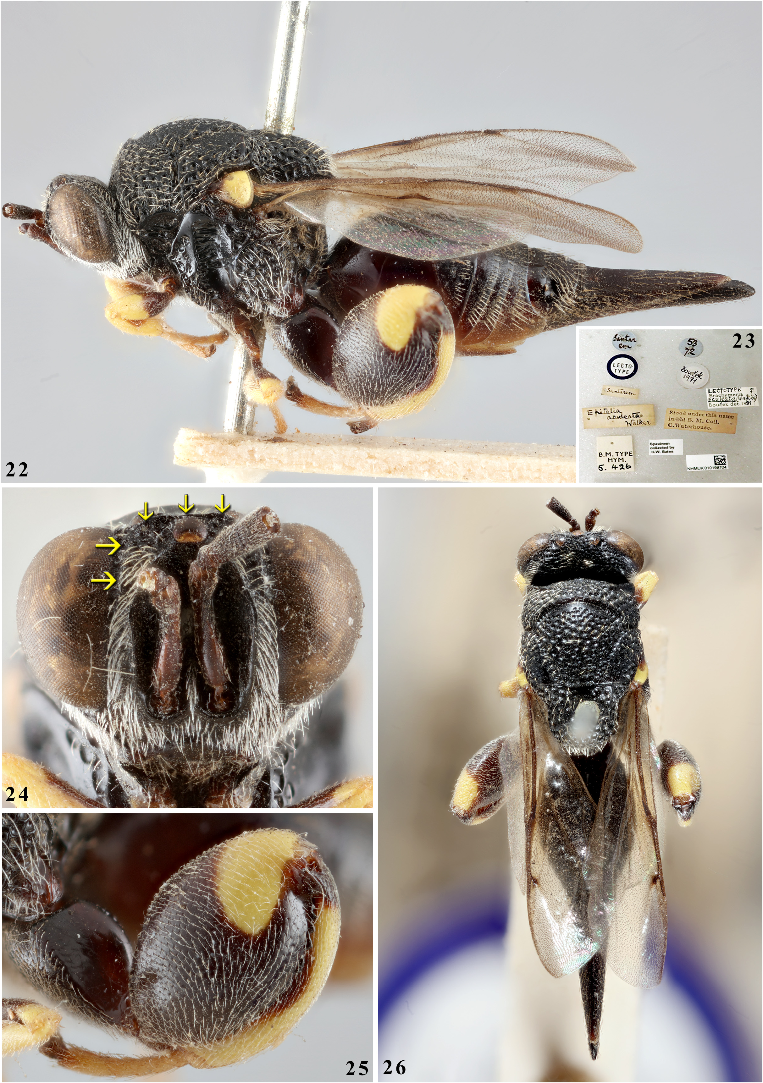
Remarks. Using the key of Burks (1960) to species of the subgenus Pseudobrachymeria , the Indian female keys to Brachymeria ( Pseudobrachymeria) laetiliae ) in having similar colour and sculpture, but although the female syntergum is acuminate it is not conspicuously elongate ( Figs 17, 20 View FIGURES 17–21 ). The Indian female also resembles B aculeata (Walker) ( Figs 22, 24–26 View FIGURES 22–26 ) in general appearance, but the latter has the hind tibia more extensively yellow with only basal and ventromesal brown patches ( Fig. 25 View FIGURES 22–26 ); further, the vertex has a transverse carina posterior to the median ocellus in addition to vertical carinae on the frons so the head has a complete, inverted U-shaped carina ( Fig. 24 View FIGURES 22–26 ).
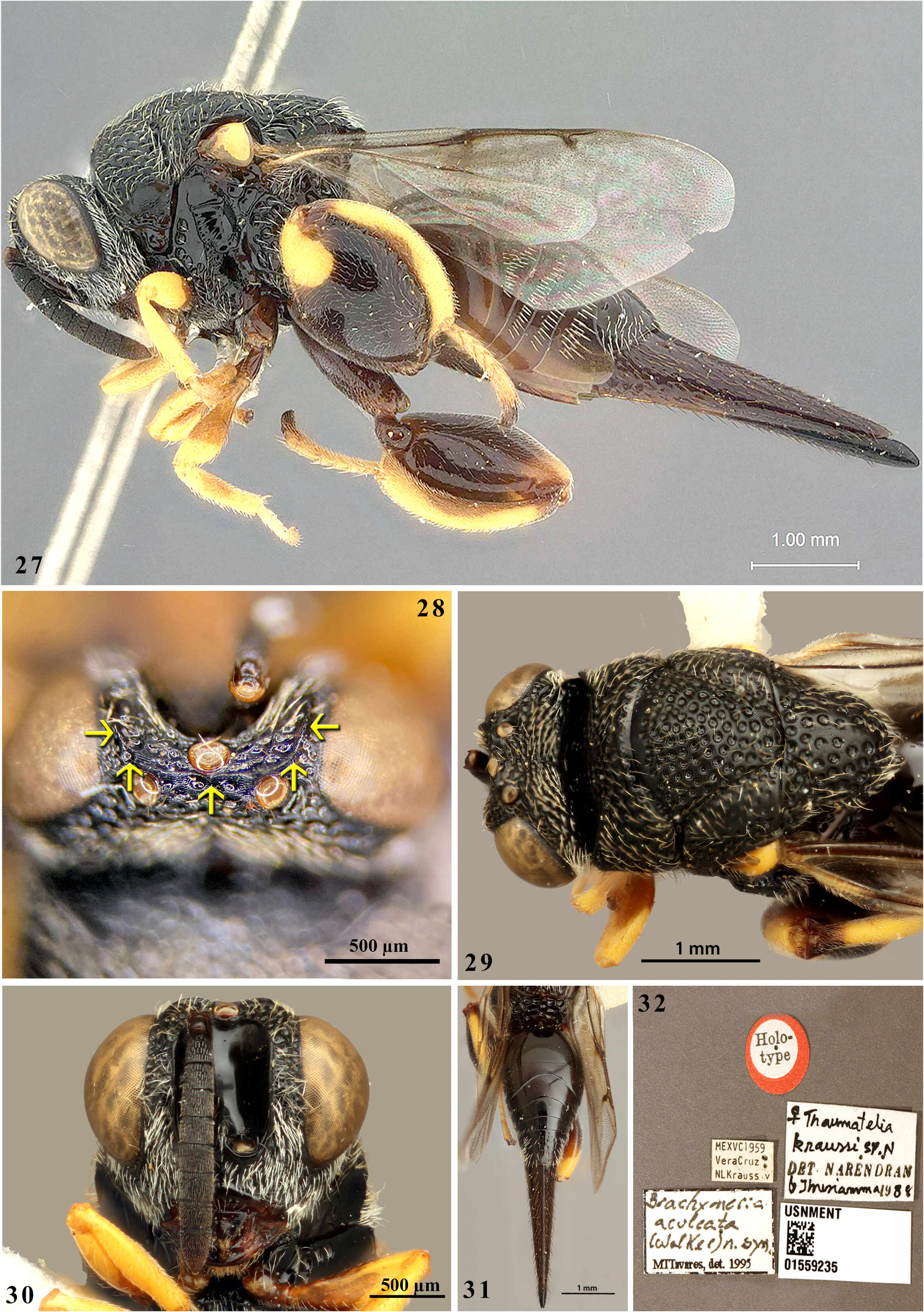
Type images of the holotype of B. kraussi (Narendran & Varghese) ( Figs 27‒31 View FIGURES 27–32 ) are similar to those of the lectotype of B. aculeata ( Figs 22, 24‒26 View FIGURES 22–26 ), and the latter bears a label by M.T. Tavares dated 1995 suggesting that B. kraussi is junior synonym of B. aculeata , though this proposed synonymy has never been validated in the literature. Present comparison of the types images of B. kraussi and B. aculeata substantiate that both have a similar hind tibia colour pattern ( Figs 22, 25 View FIGURES 22–26 , 27 View FIGURES 27–32 ) and an inverted U-shaped carina on the head ( Figs 24 View FIGURES 22–26 , 28 View FIGURES 27–32 ). However, the holotype of B. kraussi differs from the lectotype of B. aculeata in having subparallel scrobal margins ( Fig. 30 View FIGURES 27–32 ) rather than curved margins that converge dorsally toward the median ocellus ( Fig. 24 View FIGURES 22–26 ), the syntergum 0.8× the combined length of the preceding metasomal terga ( Figs 27, 31 View FIGURES 27–32 ) compared to 0.6× the combined length of the preceding metasomal terga ( Figs 22, 26 View FIGURES 22–26 ), and the mesoscutum with large round punctures well separated by sculptured interspaces ( Fig. 29 View FIGURES 27–32 ) compared to the mesoscutum being rugose-punctate with very narrow interspaces ( Fig. 26 View FIGURES 22–26 )). Hence, we do not formally synonymize the two names at this time.
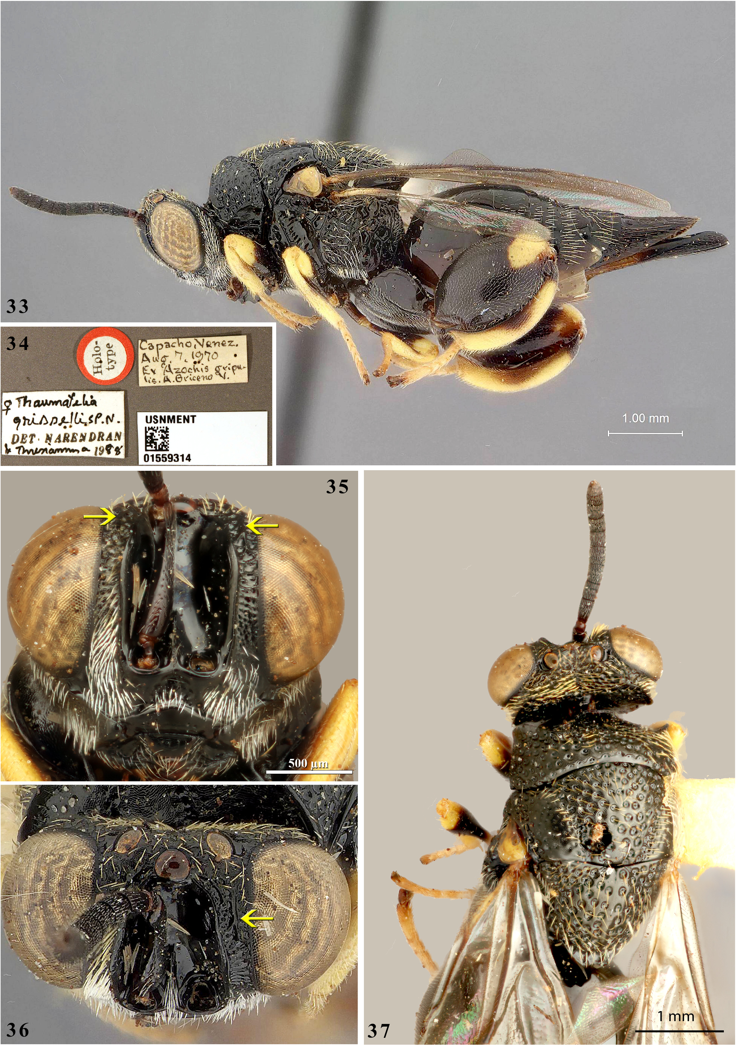
Brachymeria trinidadensis is differentiated from B. grisselli ( Figs 33, 35–37 View FIGURES 33–37 ) in having a much longer syntergum (0.7× vs. 0.4× as long as the combined length of the preceding metasomal terga); hind tibia reddishbrown with subbasal and apical yellow patch ( vs. hind tibia yellow with basal and ventromedial brown patch, Fig. 33 View FIGURES 33–37 ); mesonotum with a different sculpture pattern ( cf. Figs 12 View FIGURES 6–16 , 29 View FIGURES 27–32 ); and fore wings hyaline ( vs. fore wings infumate, Figs 33, 37 View FIGURES 33–37 ).
Brachymeria trinidadensis is differentiated from B. producta ( Figs 38, 40–43 View FIGURES 38–43 ) in having a much longer syntergum (0.7× vs. 0.4× as long as the combined length of the preceding metasomal terga and 3.96× vs. 1.64× as long as Gt 6); hind tibia reddish-brown with subbasal and apical yellow patch ( vs. hind tibia yellow with medial brown patch not extending dorsally); cercus on syntergum much closer to anterior margin ( vs. cercus on syntergum near half length, Fig. 38 View FIGURES 38–43 ).
Brachymeria trinidadensis is differentiated from B. westwoodi (Bouček, 1992, figs 76–78) in having eyes glabrous ( vs. eye sparsely setose); spiracle of Gt 6 normal ( vs. spiracle of Gt 6 raised, produced backwards); hind femur ventrally with nine well-separated irregular teeth ( vs. hind femur ventrally with 13 well separated teeth).
Brachymeria trinidadensis is differentiated from B. bicolor ( Girault, 1912) (Bouček, 1992, fig. 75) in having spiracle of Gt 6 normal ( vs. spiracle of Gt 6 placed dorsally on a raised hump); hind femur ventrally with nine wellseparated irregular teeth ( vs. hind femur ventrally with eight teeth); hind tibia reddish-brown with subbasal and apical yellow patch ( vs. hind tibia yellow with median brownish black patch).
Brachymeria trinidadensis is differentiated from B. pyramidea ( Bouček & Delvare 1992, figs 60–65) in having hind femur ventrally with nine well-separated irregular teeth ( vs. hind femur ventrally with 14 teeth); hind tibia reddish-brown with subbasal and apical yellow patch, ( vs. hind tibia dorsally (except base) yellow); wings hyaline ( vs. wings subinfumate); notauli shallow ( vs. notauli unusually deep); syntergum 4.1× Gt 6 ( vs. syntergum 1.9× Gt 6).


| PMV |
Provincial Museum |
No known copyright restrictions apply. See Agosti, D., Egloff, W., 2009. Taxonomic information exchange and copyright: the Plazi approach. BMC Research Notes 2009, 2:53 for further explanation.
|
Kingdom |
|
|
Phylum |
|
|
Class |
|
|
Order |
|
|
Family |
|
|
Genus |
Brachymeria trinidadensis (Narendran & Varghese)
| Binoy, C., Santhosh, S. & Nasser, M. 2022 |
Thaumatelia trinidadensis
| Halstead, J. A. 1991: 952 |
| Narendran, T. C. & Varghese, T. 1989: 47 |