
Proschizorhynchella shibazakii, Takeda & Kajihara, 2018
|
publication ID |
https://doi.org/10.12782/specdiv.23.1 |
|
publication LSID |
lsid:zoobank.org:pub:A9B333A2-FD81-476A-A805-D280C2194964 |
|
DOI |
https://doi.org/10.5281/zenodo.5527060 |
|
persistent identifier |
https://treatment.plazi.org/id/EC658D8C-E9ED-4677-B119-0A84758174CE |
|
taxon LSID |
lsid:zoobank.org:act:EC658D8C-E9ED-4677-B119-0A84758174CE |
|
treatment provided by |
Felipe |
|
scientific name |
Proschizorhynchella shibazakii |
| status |
sp. nov. |
Proschizorhynchella shibazakii View in CoL sp. nov.
( Figs 10–14 View Fig View Fig View Fig View Fig View Fig ; Table 1 View Table 1 )
Material examined. Holotype: ICHUM 4275 View Materials , adult, whole mount, 43°12′33″N, 140°51′31″E, Oshoro , Hokkaido, Japan, intertidal sand, 13 June 2011 GoogleMaps . Paratypes: ICHUM 4276–4278, three adults, whole mounts, same data as holotype; ICHUM 4279, 4280, two adults, serial sagittal sections, same data as holotype; ICHUM 4281, 4282, two adults, serial transverse sections, same data as holotype; ICHUM 4283, one adult, whole mount, type locality, 21 May 2012; ICHUM 4861, egg, whole mount, laid by animals collected on 1 July 2013.
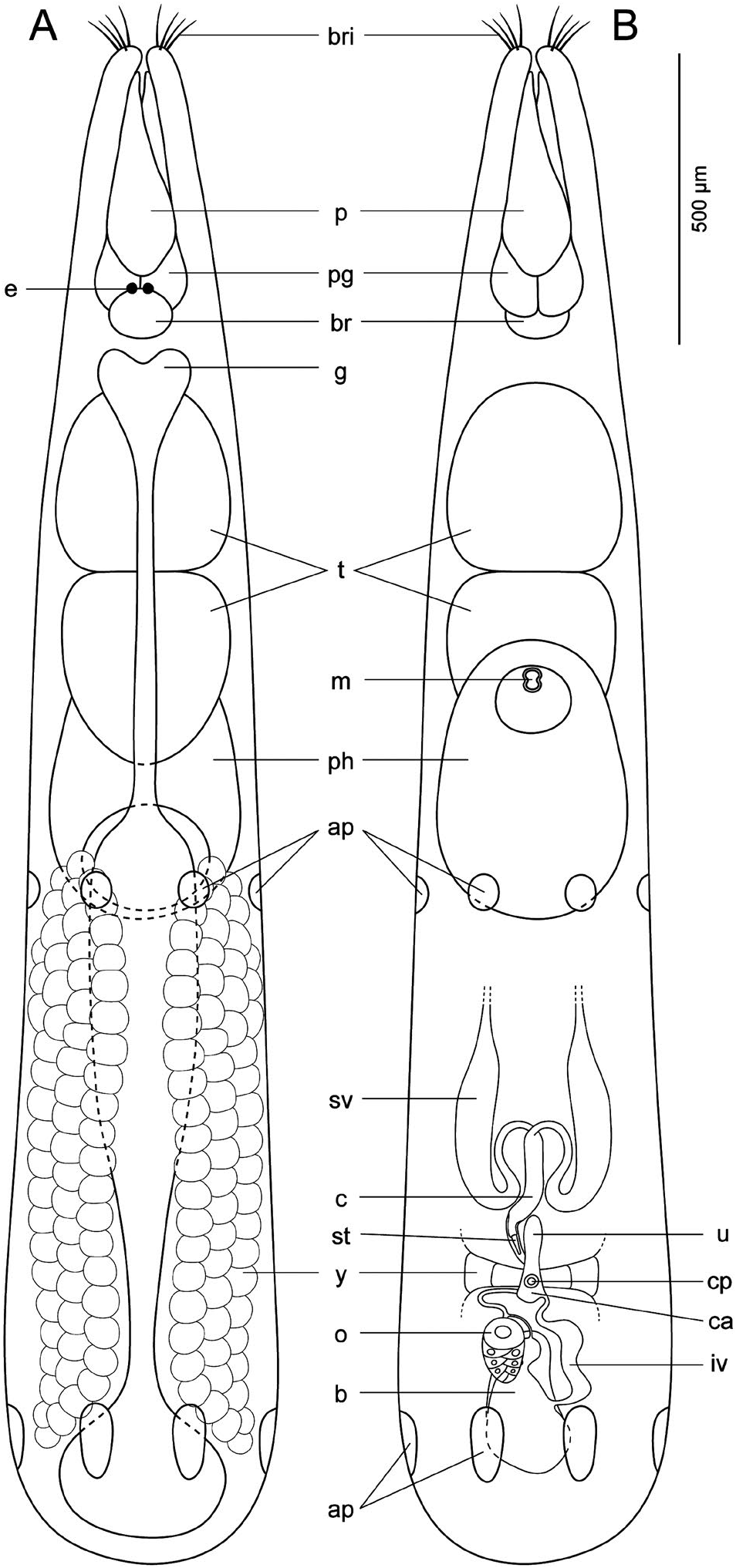
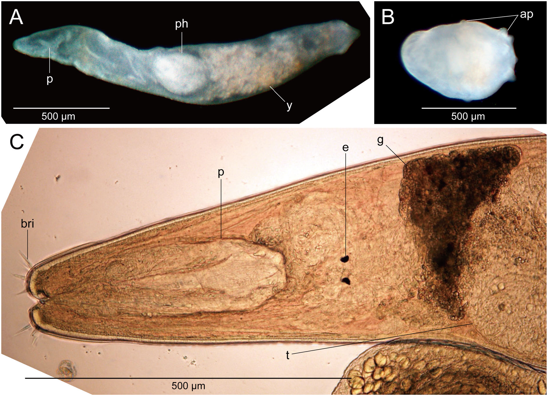
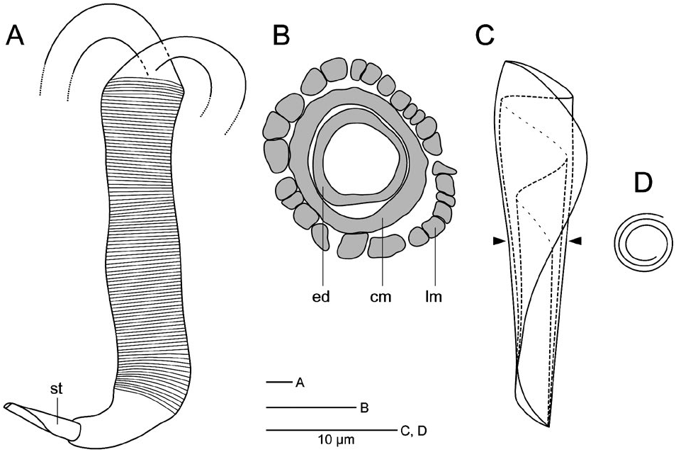
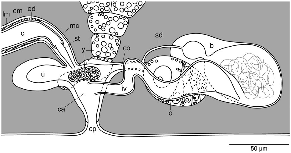
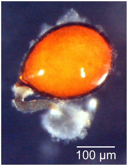
Description. Living animal body approximately 2.6 mm long and 0.5 mm wide ( Figs 10 View Fig , 11A View Fig ). Four pairs of bristles located at slender anterior tip of body ( Figs 10 View Fig , 11C View Fig ). Proboscis 350 µm long, 120 µm wide; pair of proboscis glands 140 µm long, 80 µm wide ( Fig. 10 View Fig ). Pair of black eyes situated anterior to brain ( Figs 10 View Fig , 11C View Fig ). Gut anteroposteriorly elongated. Two testes 300–330 µm in diameter ( Figs 10 View Fig , 11C View Fig ). Pair of yolk glands 1 mm long, 180 µm wide ( Figs 10 View Fig , 11A View Fig ). Pharynx 480 µm long, 330 µm wide ( Figs 10 View Fig , 11A View Fig ). Two adhesive girdles present; anterior one located at level of posterior end of pharynx, posterior one near caudal end; each girdle comprised of six adhesive papillae arranged in regular intervals ( Figs 10 View Fig , 11B View Fig ). Pair of seminal vesicles, each 620 µm long, 60 µm wide, located posterior to pharynx ( Fig. 10 View Fig ). Male copulatory organ tubular in shape, 240 µm long, 30 µm wide, with ejaculatory duct surrounded by circular muscles and further surrounded by longitudinal muscles ( Figs 10 View Fig , 12A, B View Fig , 13 View Fig ); copulatory organ tapering toward its tip, equipped with stylet and situated in male genital canal. Stylet cone shaped, 29–31 µm long ( 31 µm in holotype), 7 µm wide, comprised of thin sclerotic sheet rolled up three times ( Fig. 12C, D View Fig ). Male genital canal opens to anterodorsal part of common atrium of the latter ( Fig. 13 View Fig ). Uterus 90 µm long, 30 µm wide, anterior to common atrium ( Figs 10 View Fig , 13 View Fig ). Each yolk gland connected to each side of common atrium ( Figs 10 View Fig , 13 View Fig ). Common genital pore opening on ventral side of body between two adhesive girdles ( Figs 10 View Fig , 13 View Fig ). Ovary 110 µm long, 70 µm wide, anteriorly connected to posterodorsal portion of common atrium via a common oviduct ( Figs 10 View Fig , 13 View Fig ). Bursa oval in dorsal view, 250 µm long, 150 µm wide; bursal tissue divided into two (smaller anterior and larger posterior) parts by constriction; spermatids observed in posterior bursal tissue in all specimens observed; anterior bursal tissue leading forward to connect to common oviduct near ovary via narrow sperm duct ( Figs 10 View Fig , 13 View Fig ). Egg oval, 260 µm long, 200 µm wide, covered in brown shell with colorless axis ( Fig. 14 View Fig ).
Etymology. The specific name is a noun in the genitive case, derived from the name Mr. Kouji Shibazaki, a caretaker of Oshoro Marine Station, Hokkaido University.
Remarks. Proschizorhynchella shibazakii can be distinguished from all congeners based on the characteristics listed in Table 1 View Table 1 except P. papillata . These two species, however, can be distinguished based on the shape of the male copulatory organ. The differences in morphological characteristics between P. shibazakii sp. nov. and P. papillata are ( i) the number of the apical sensory bristles, which is eight in P. shibazakii and four in P. papillata ; ( ii) the male copulatory organ, which is narrow and tubular in P. shibazakii , and bulb shaped in P. papillata ; ( iii) the internal part of the circular muscles, which is thin in P. shibazakii but thick in P. papillata ; ( iv) the border cells which are present in P. shibazakii but absent in P. papillata ; and ( v) the length of the stylet, which is 29–31 µm ( 31 µm in holotype) in P. shibazakii and 55–57 µm in P. papillata . Proschizorhynchella shibazakii cannot be distinguished from P. caudociliata based on the characteristics listed in Table 1 View Table 1 ; however, they differ in the structure of the copulatory stylet (see Remarks for P. caudociliata ).
| ICHUM |
Invertebrate Collection of the Hokkaido University Museum |
No known copyright restrictions apply. See Agosti, D., Egloff, W., 2009. Taxonomic information exchange and copyright: the Plazi approach. BMC Research Notes 2009, 2:53 for further explanation.
|
Kingdom |
|
|
Phylum |
|
|
Class |
|
|
Order |
|
|
SubOrder |
Kalyptorhynchia |
|
Family |
|
|
Genus |