
Tiphia ( Tiphia ) mediocarinata Han, Chen & Li, 2023
|
publication ID |
https://doi.org/10.11646/zootaxa.5284.1.1 |
|
publication LSID |
lsid:zoobank.org:pub:10739869-526E-4B61-A955-901724CA7198 |
|
DOI |
https://doi.org/10.5281/zenodo.7921186 |
|
persistent identifier |
https://treatment.plazi.org/id/7322004E-737E-FFAE-5A82-470F0C8AA08C |
|
treatment provided by |
Plazi |
|
scientific name |
Tiphia ( Tiphia ) mediocarinata Han, Chen & Li |
| status |
sp. nov. |
Tiphia ( Tiphia) mediocarinata Han, Chen & Li , sp. nov.
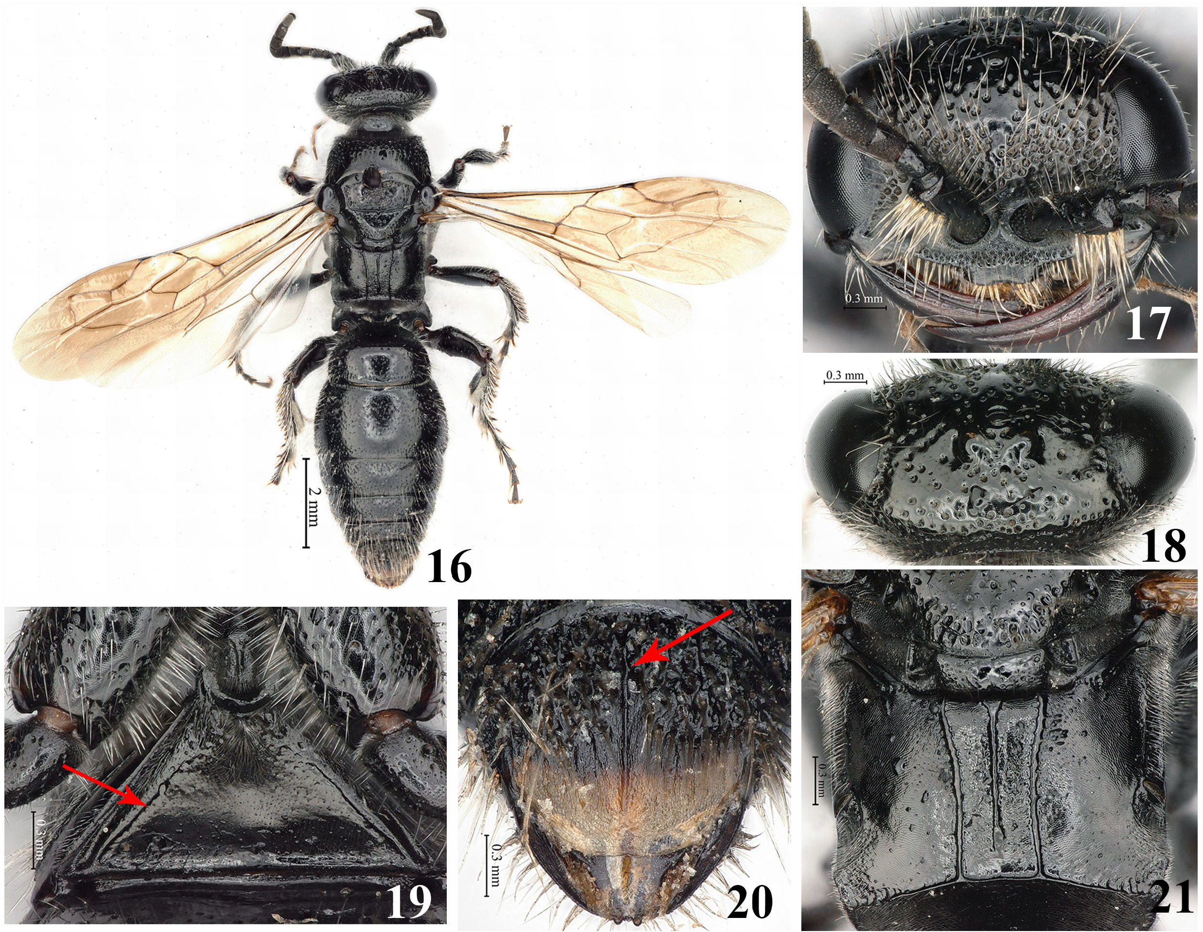
( Figs 16–21 View FIGURES 16–21 )
Material examined. Holotype, ♀, China, Yunnan prov., Dehong City, Yingjiang County, Nabang Town , 24°45′26″N, 97°33′50″E, 221 m, 18.VIII.2017, Pan Huang ( CNU). GoogleMaps
Diagnosis. This species can be recognized by the following combination of characters: horizontal area of propodeal dorsal face ( Fig. 21 View FIGURES 16–21 ) with a few sparse punctures, oblique area impunctate and without submarginal carina, posteriorly with a few short longitudinal striae connecting transverse carina; S1 ( Fig. 19 View FIGURES 16–21 ) anteriorly with some irregular pits, medially with sparse minute punctures; pygidium ( Fig. 20 View FIGURES 16–21 ) with a medial longitudinal carina; inner face of hind basitarsus medially with longitudinal groove.
Description. Female ( Fig. 16 View FIGURES 16–21 ). Body length 12.1 mm, forewing length 8.1 mm.
Color. Body almost black, with pale brown setae; mandible, tegula, pterostigma, veins and legs dark brown; posterior half of pygidium ( Fig. 20 View FIGURES 16–21 ) brown; wings ( Fig. 16 View FIGURES 16–21 ) infuscate.
Head. Mandible ( Fig. 17 View FIGURES 16–21 ) without distinct medial transverse groove and preapical denticle, atmost with obsolete intermittent impressed line; clypeus ( Fig. 17 View FIGURES 16–21 ) basally with dense punctures and apically impunctate, apex medially emarginated; W: OW: L: IOD=10: 4.8: 4.9: 5.9; OOD: POD: Od=10: 5.9: 3.3; AOD: WAS: IAD: CL: CAW=10: 4.6: 3.2: 6.2: 6.7; lower frons ( Fig. 17 View FIGURES 16–21 ) with medial longitudinal narrow groove and dense punctures, upper frons with sparse punctures; vertex ( Fig. 18 View FIGURES 16–21 ) with sparse punctures, interspaces smooth.
Mesosoma . Pronotal anterior carina ( Fig. 16 View FIGURES 16–21 ) absent; anterior half of pronotum with big punctures evenly distributed, posterior half impunctate; pronotum latero-ventrally with distinct groove in middle, area above groove smooth, area below groove with dense oblique striae; mesoscutum ( Fig. 16 View FIGURES 16–21 ) medially with coarse and dense punctures, sparser laterally, anterior medial groove separated from notaulus; mesopleuron with dense punctures; mesoscutellum posteriorly with irregular and dense punctures; metanotum ( Fig. 21 View FIGURES 16–21 ) with several sparse punctures mixed with dense minute punctures; horizontal area of propodeal dorsal face ( Fig. 21 View FIGURES 16–21 ) with a few sparse punctures, oblique area impunctate and without submarginal carina, posteriorly with a few short longitudinal striae connecting transverse carina; propodeal areola ( Fig. 21 View FIGURES 16–21 ) rectangular, APWL=5.9: 4.9: 11.2, medial longitudinal carina reaching posterior 4/5 of areola; lateral surface of propodeum dorsally with dense long oblique wrinkles, ventrally impunctate; posterior surface of propodeum with dense punctures and complete medial longitudinal carina; tegula atmost slightly longer than middle width, posterior margin without transverse impressed line; second intercubital vein of forewing ( Fig. 16 View FIGURES 16–21 ) sinuous; fore tibia with one spur apically, mid and hind tibiae with 2 spurs apically; inner face of hind basitarsus medially with longitudinal groove.
Metasoma. Anterior half of T1 ( Fig. 16 View FIGURES 16–21 ) with sparse shallow punctures; subposterior area of T1 close to apex and with one transverse row of punctures; S1 ( Fig. 19 View FIGURES 16–21 ) anteriorly with some irregular pits, medially with sparse minute punctures, and with posterolateral groove and approaching the anterior apex; T2 antero-laterally with costate transverse depression ( Fig. 16 View FIGURES 16–21 ), medially obsolete; T2–T4 with sparse punctures; S2–S5 with sparser punctures than T2–T4; pygidium ( Fig. 20 View FIGURES 16–21 ) with a medial longitudinal carina, anterior half with coarse and dense punctures, strongly reticulate, and posterior half impunctate and coriaceous.
Male. Unknown.
Distribution. China ( Yunnan).
Etymology. The specific name mediocarinata is derived from the two latin words: medius (= medial) + carinata (= carinate), referring to pygidium with a medial longitudinal carina.
| CNU |
Capital Normal University, College of Life Sciences |
No known copyright restrictions apply. See Agosti, D., Egloff, W., 2009. Taxonomic information exchange and copyright: the Plazi approach. BMC Research Notes 2009, 2:53 for further explanation.
|
Kingdom |
|
|
Phylum |
|
|
Class |
|
|
Order |
|
|
Family |
|
|
Genus |
|
|
SubGenus |
Tiphia |