
Geniatini, Burmeister, 1844
|
publication ID |
https://doi.org/10.11646/zootaxa.874.1.1 |
|
publication LSID |
lsid:zoobank.org:pub:1EAA2669-E530-4F6D-901B-7B565C1F5301 |
|
DOI |
https://doi.org/10.5281/zenodo.10532342 |
|
persistent identifier |
https://treatment.plazi.org/id/8B4A3E15-FFAA-5122-AA5C-46B8FABAD719 |
|
treatment provided by |
Felipe |
|
scientific name |
Geniatini |
| status |
|
Key to the Genera of Geniatini View in CoL
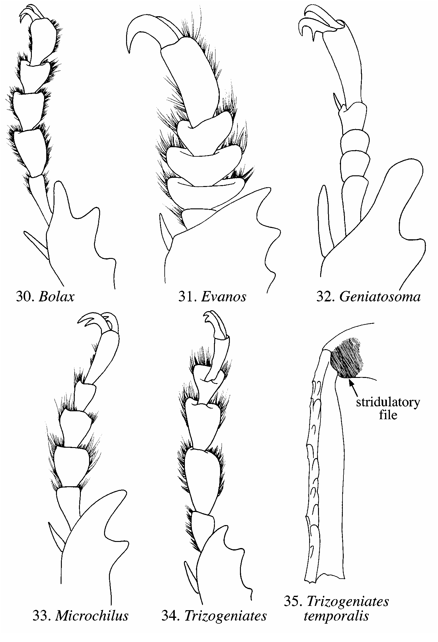
Males: Protarsomeres dorsoventrally flattened, densely setose ventrally ( Figs. 30–31, 33– 34 View FIGURES 30–35 ); terminal sternite with margin emarginated; abdominal sternites in lateral view appearing concave or flat. Females: Protarsomeres dorsoventrally flattened or not, with or without dense ventral setae; terminal sternite with margin entire or rounded, not emarginated; abdominal sternites in lateral view appearing convex.
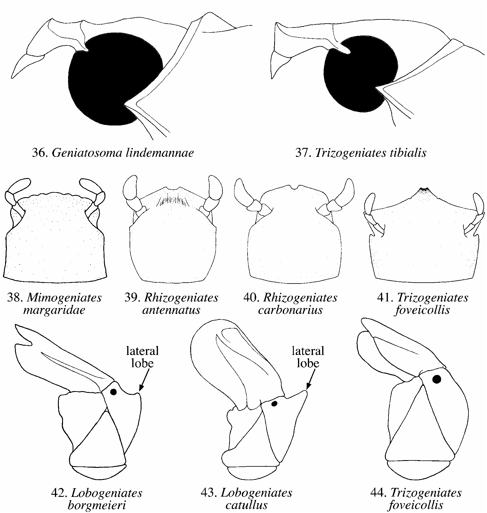
1. Mentum with apicomedial, toothlike projection ( Fig. 16 View FIGURES 14–18 , 41 View FIGURES 36–44 ) .................................... 3
1’. Mentum without apicomedial, toothlike projection ( Figs. 38–40 View FIGURES 36–44 ) ............................. 2
2. Apex of mentum with medial notch, not crenulate ( Figs. 39–40 View FIGURES 36–44 ). All claws simple on all legs........................................................................................... Rhizogeniates Ohaus
2’. Apex of mentum crenulate ( Fig. 38 View FIGURES 36–44 ). Modified claw moderately split on all legs ....... .................................................................................................. Mimogeniates Martínez
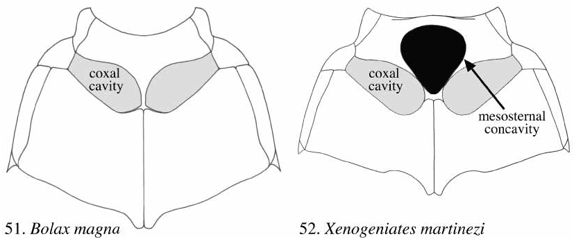
3. Mesosternum anterior to mesocoxae strongly concave ( Fig. 52 View FIGURES 51–52 ) .................................................................................................................... Xenogeniates Villatoro & Jameson
3’. Mesosternum anterior to mesocoxae flat or slightly convex, not strongly concave ( Fig. 51 View FIGURES 51–52 ) ............................................................................................................................... 4
4. Stipes of maxilla produced, with welldeveloped lateral lobe ( Fig. 43 View FIGURES 36–44 ) or lateral angle ( Fig. 42 View FIGURES 36–44 )......................................................................................... Lobogeniates Ohaus
4’. Stipes of maxilla not produced, instead rounded or broadly rounded ( Fig. 44 View FIGURES 36–44 ) ........... 5
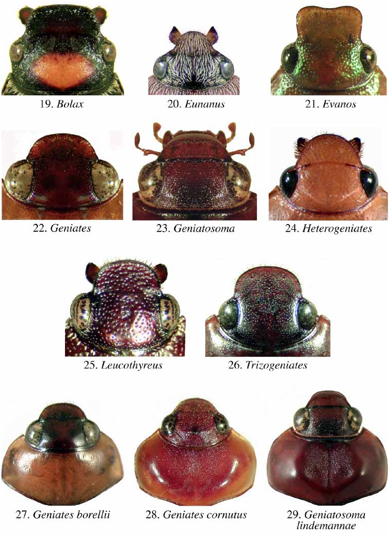
5. Mandible with rounded, recurved, apical lobe ( Fig. 20 View FIGURES 19–29 ). Dorsal surface with abundant, decumbent, white setae. Antennal club of male twice length of segments 2–7; antennal club of female subequal to segments 2–7 ....................................... Eunanus Ohaus
5’. Mandible lacking rounded, recurved, apical tooth; instead simple ( e.g., Fig. 19 View FIGURES 19–29 ). Dorsal surface with or without sparse setae. Antennal club of male and female subequal to or slightly longer than segments 2–7 ....................................................................... 6
6. Length of antennal club half or less than half length of first antennal segment ( Figs. 5 View FIGURES 5–8 , 23 View FIGURES 19–29 ). Clypeal apex (in lateral view) sloped 45º with respect to dorsal plane of clypeus ( Figs. 23 View FIGURES 19–29 , 36 View FIGURES 36–44 ). Male tarsomeres simple, not flattened and dilated ( Fig. 32 View FIGURES 30–35 ) .................................................................................................................. Geniatosoma Costa Lima
6’. Length of antennal club more than half length of first antennal segment. Clypeal apex (in lateral view) sloped 60–90º with respect to dorsal plane of clypeus ( Fig. 37 View FIGURES 36–44 ). Male tarsomeres dorsoventrally flattened and dilated ( e.g., Fig. 34 View FIGURES 30–35 ) .................................... 7
7. Form of clypeus parabolic, apex not reflexed ( Fig. 24 View FIGURES 19–29 ). Mandible exposed, apex narrowly rounded ( Fig. 24 View FIGURES 19–29 ). Male with all claws appearing simple on all legs ....................................................................................................................... Heterogeniates Ohaus
7’. Form of clypeus not parabolic (instead rounded, quadrate), apex reflexed ( e.g., Figs. 19–20, 22, 25–26 View FIGURES 19–29 ). Mandible exposed or not, apex broadly rounded ( e.g., Figs. 19, 23, 25 View FIGURES 19–29 ). Male with claws obviously toothed on some or all legs ...................................... 8
8. Length of protarsomeres 2–4 subequal in length to protarsomere 5 ( Fig. 31 View FIGURES 30–35 ). Clypeus of male with lateral margins expanded, apex quadrate ( Fig. 21 View FIGURES 19–29 ); clypeus of female with lateral margins parallel, apex quadrate............................................ Evanos Ohaus
8’. Length of protarsomeres 2–4 greater than length of protarsomere 5 ( Figs. 30, 33–34 View FIGURES 30–35 ). Clypeus of male and female with lateral margins constricted, apex rounded or trapezoidal ( e.g., Figs. 19, 22, 25–28 View FIGURES 19–29 ) .................................................................................. 9
9. Elytral margin with deep, setose punctures on lateral edge from apex of metepisternum to apex of elytra ( Figs. 45a–b, 46a–b View FIGURES 45–50 )......................................................................... 10
9’. Elytral margin without deep, setose punctures on lateral edge from apex of metepisternum to apex of elytra ................................................................................................. 11
10. Elytral margin with welldeveloped stridulatory ridge and with rigid stridulatory setae ( Fig. 45a–b View FIGURES 45–50 ). Apex of metafemur (dorsal view) with stridulatory patch ( Fig. 35 View FIGURES 30–35 ) ................................................................................................................ Trizogeniates Ohaus
10’.Elytral margin lacking stridulatory ridge and without rigid stridulatory setae ( Fig. 46a– b View FIGURES 45–50 ). Apex of metafemur (dorsal view) lacking stridulatory patch.......... Geniates Ohaus
11. Eyes small, interocular width greater than 6 transverse eye diameters ( e.g., Fig. 19 View FIGURES 19–29 ) 12
11’. Eyes larger, interocular width less than 5 transverse eye diameters ( e.g., Fig. 25 View FIGURES 19–29 )....... ..................................................................................................... Leucothyreus Macleay
12. Protarsomere 5 dorsoventrally flattened, width more than half length ( Fig. 30 View FIGURES 30–35 ). Length of body from apex of clypeus to apex of elytra more than 9.0 mm ............................... .......................................................................................... Bolax Fischer von Waldheim
12’.Protarsomere 5 dorsoventrally flattened or not; if flattened, then width less than half length. Length of body from apex of clypeus to apex of elytra less than 9.0 mm ........ ................................................................................................... Microchilus Blanchard


No known copyright restrictions apply. See Agosti, D., Egloff, W., 2009. Taxonomic information exchange and copyright: the Plazi approach. BMC Research Notes 2009, 2:53 for further explanation.