
Andromma Simon, 1893
|
publication ID |
https://doi.org/10.5852/ejt.2022.850.1997 |
|
publication LSID |
lsid:zoobank.org:pub:E8AD897F-2076-4850-9520-BB79B1EAFFEA |
|
DOI |
https://doi.org/10.5281/zenodo.7433847 |
|
persistent identifier |
https://treatment.plazi.org/id/9C479E6B-FFF1-FFD8-FF12-F755FA90F8D5 |
|
treatment provided by |
Felipe |
|
scientific name |
Andromma Simon, 1893 |
| status |
|
Key to the species of Andromma Simon, 1893 View in CoL
Males
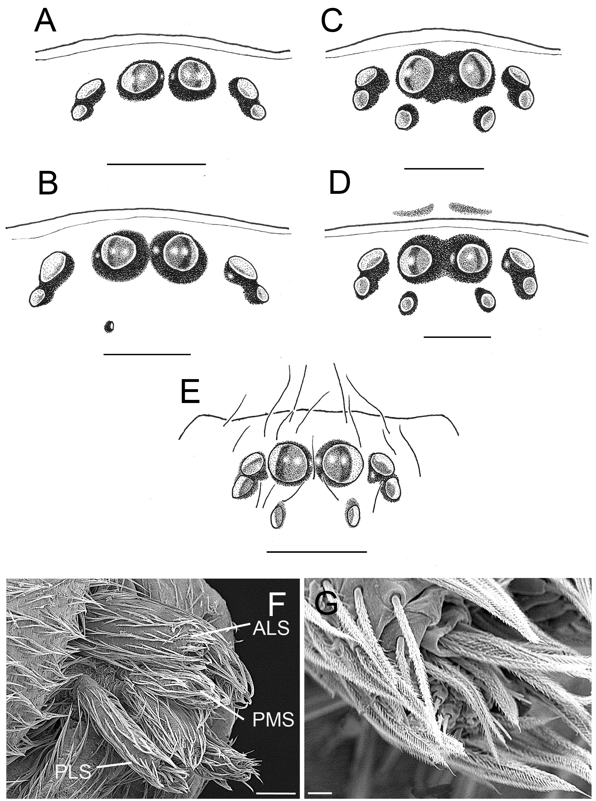
1. Posterior eyes partly or completely reduced ( Figs 2A–B View Fig , 3 View Fig )............................................................. 2
– Posterior eyes present, eight eyes ( Fig. 2C–E View Fig ) .................................................................................. 4
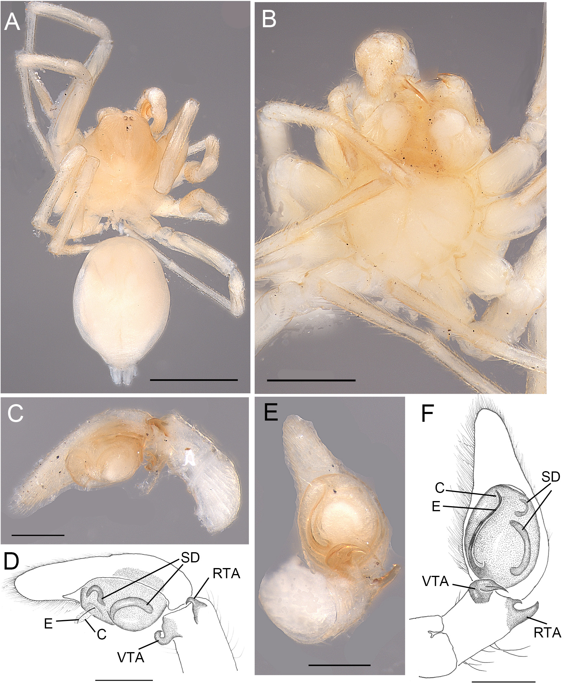
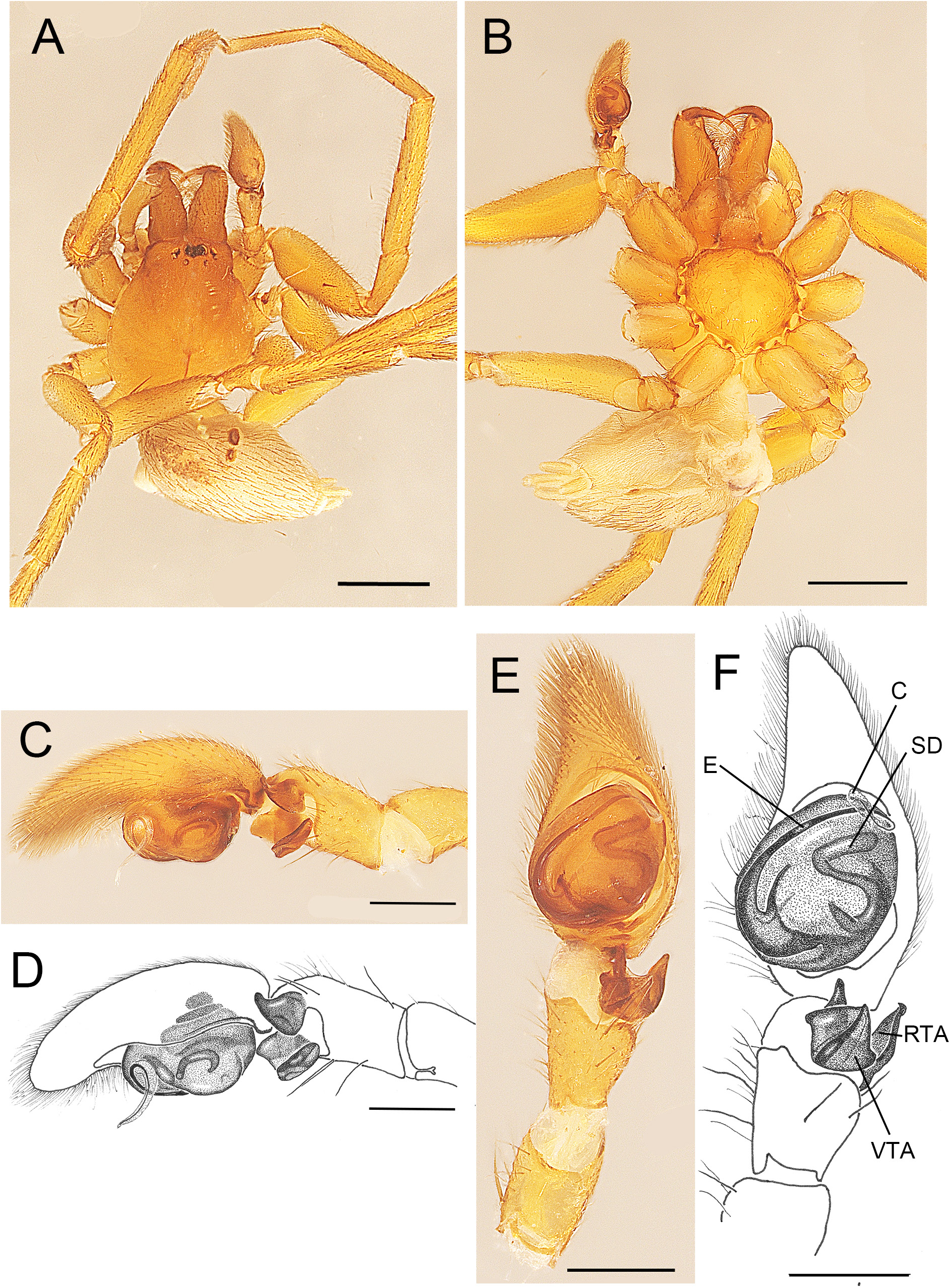
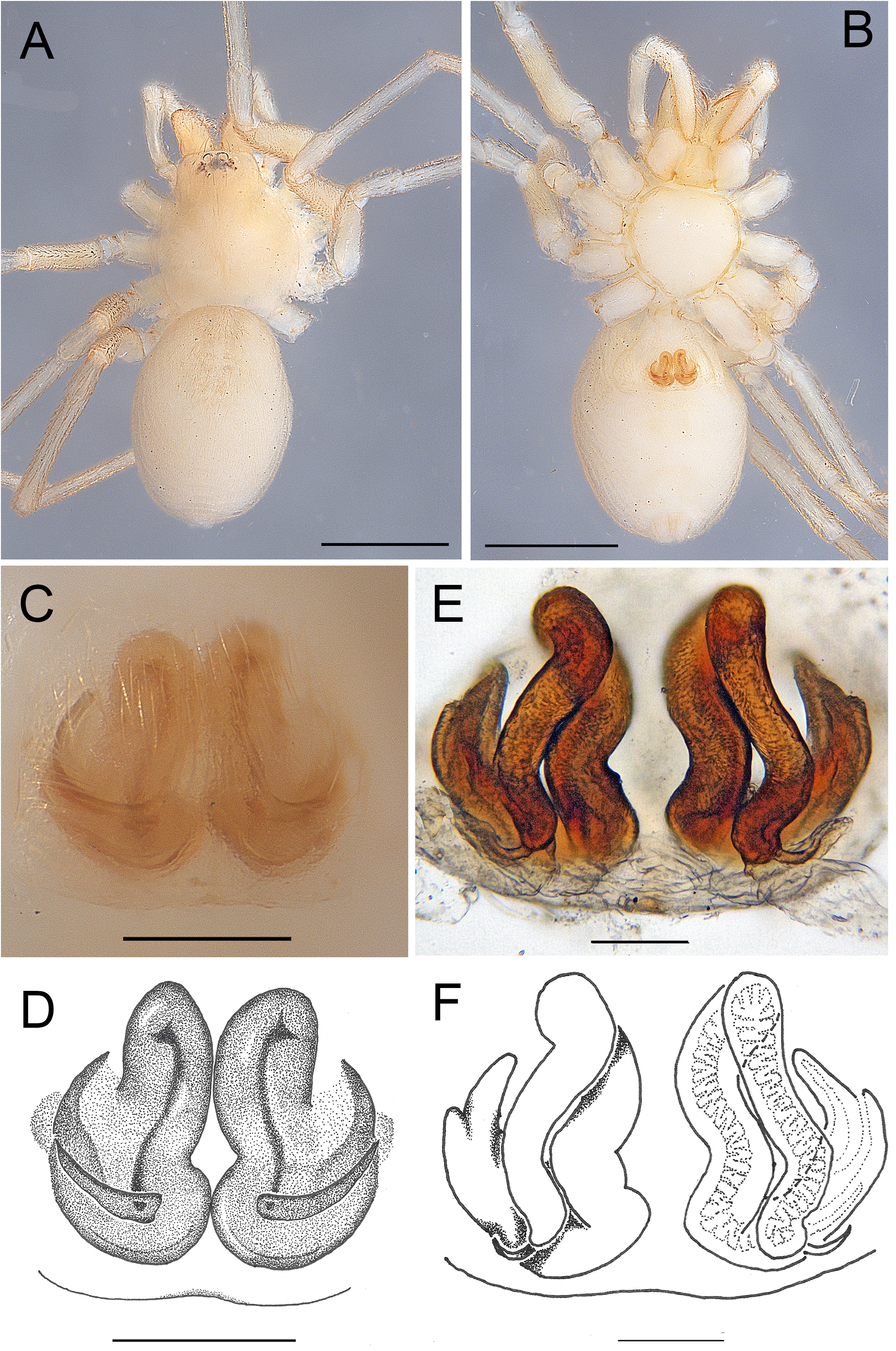
2. Animal of medium size ( 5 mm), only PME missing, RTA large and arrow-shaped ( Fig. 17C–F View Fig ) ..... .......................................................................................................................... A. deogratias sp. nov.
– Animal small ( 3 mm)......................................................................................................................... 3
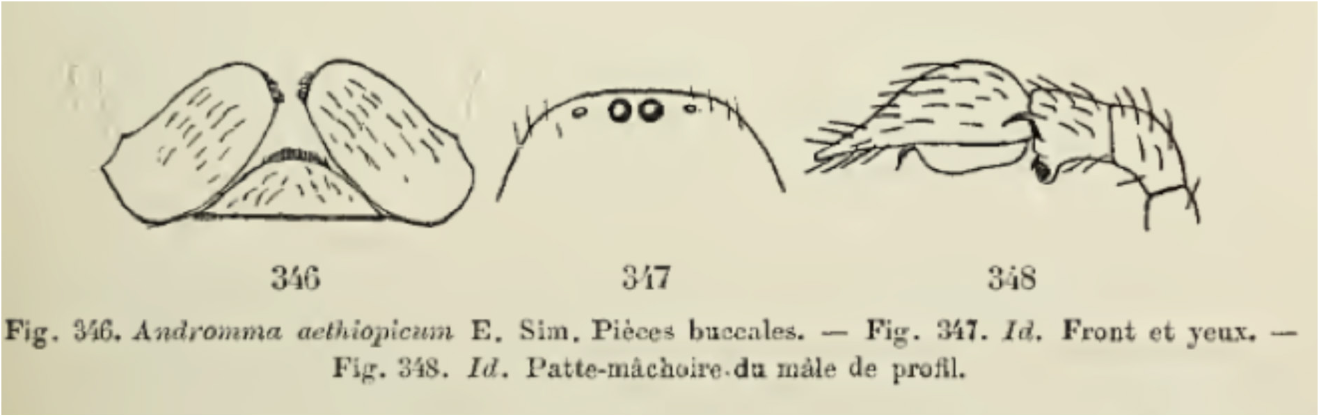
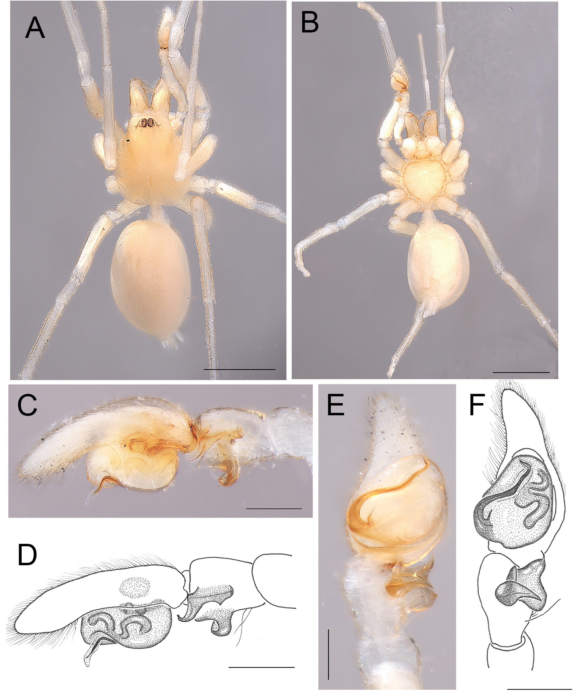
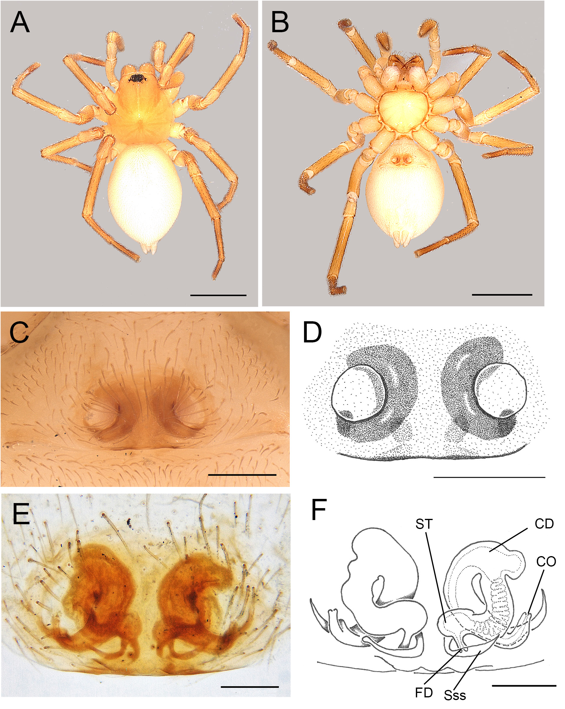
3. Sternum wider than long ( Fig. 4B View Fig ), only AME present ( Fig. 4A View Fig )......... A. aethiopicum Simon, 1893 View in CoL
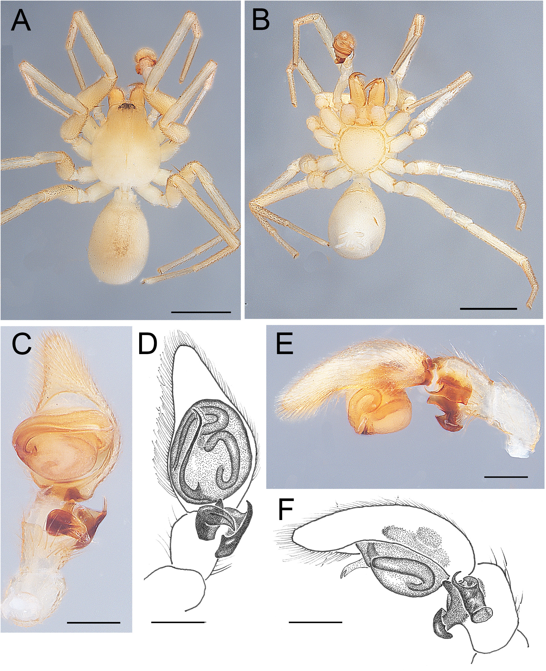
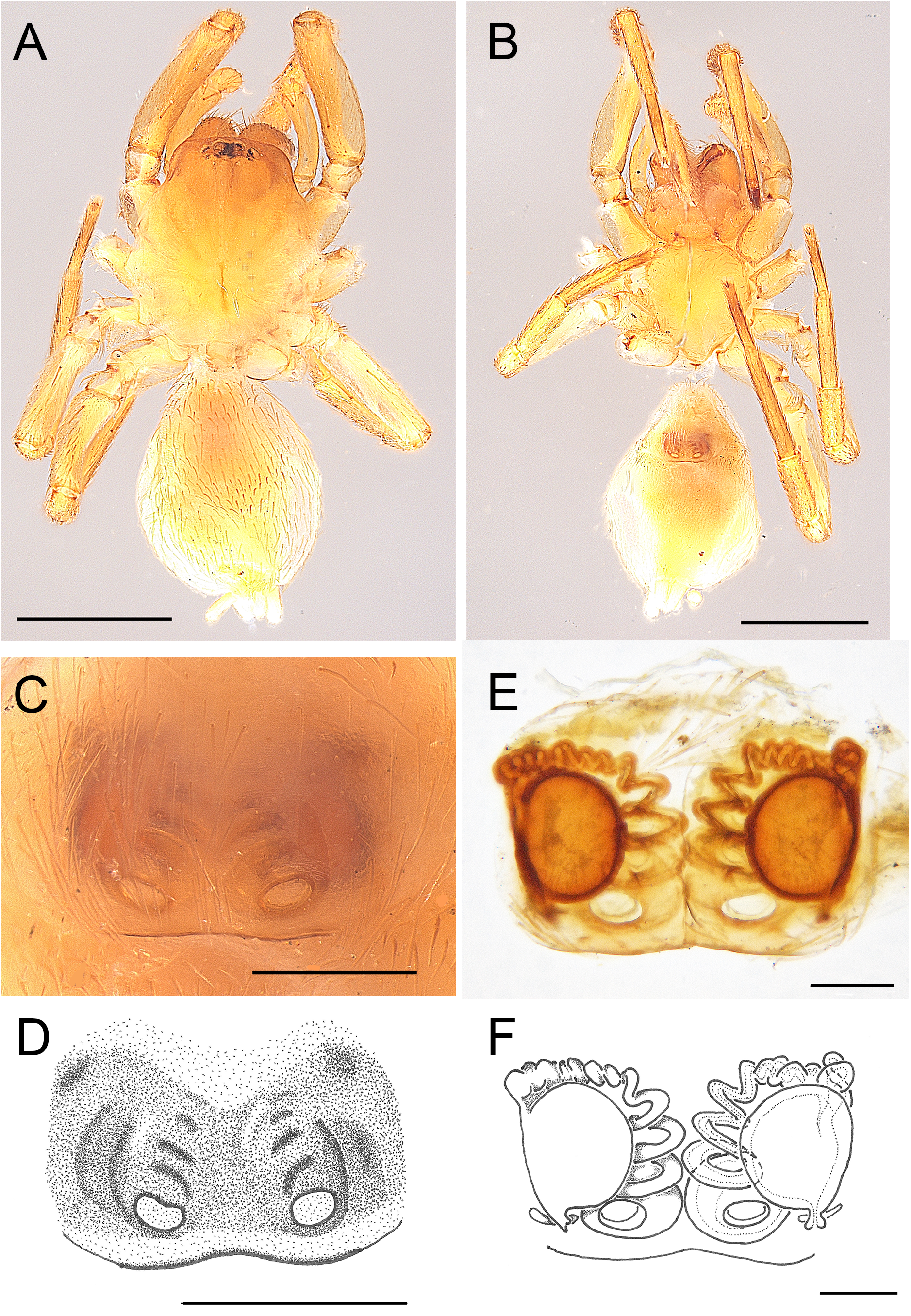
– Sternum as long as wide ( Fig. 6B View Fig ), only PME missing ( Fig. 6A View Fig ).................... A. albinovani sp. nov.
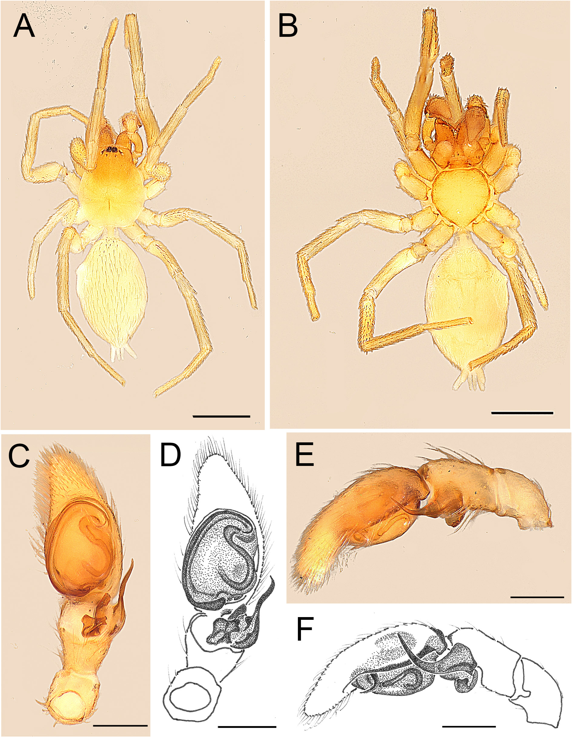
4. VTA relatively simple in ventral view: globular, heart-shaped, mushroom-shaped or hook-shaped (e.g., Figs 10E–F View Fig , 24C–D View Fig , 33E–G View Fig ).................................................................................................... 5
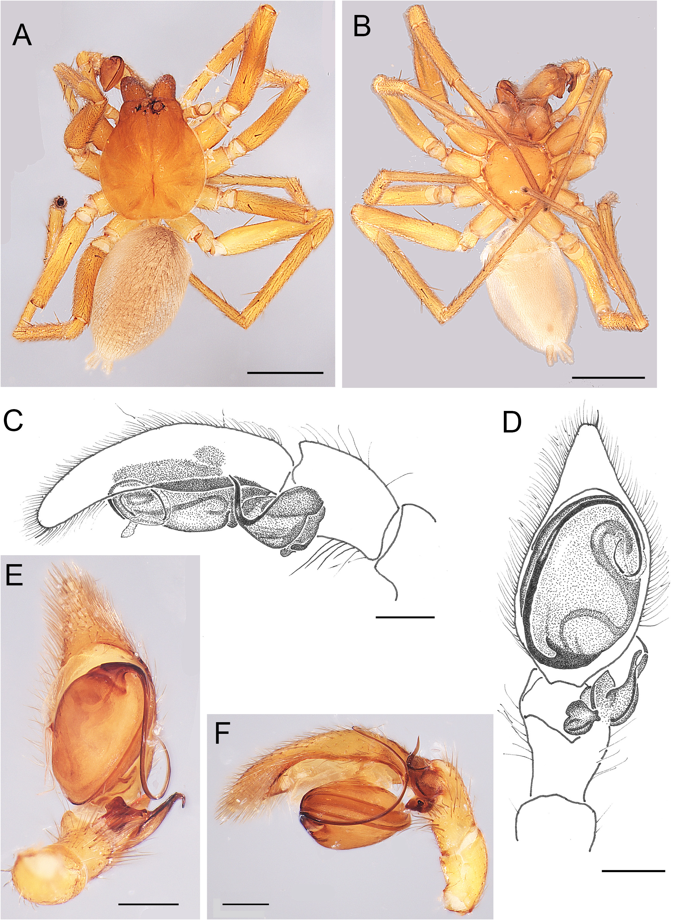
– VTA more complex in ventral view, often sail-shaped or flag-shaped ( Figs 13C–D View Fig , 21E–F View Fig , 28E–F View Fig , 36C–D View Fig ) ............................................................................................................................................ 10
5. Ventral part of RTA globular VTA subtriangular with rounded corners, embolus median in ventral view, bent in retrolateral direction, with slightly curved tip ( Fig. 24C–F View Fig )..... A. ghesquierei sp. nov.
– VTA heart-shaped, mushroom-shaped or hooked, embolus retrolateral in ventral view, bent in prolateral direction............................................................................................................................. 6
6. VTA shaped like a mushroom or a mooring post in ventral view, bifid in retrolateral view, RTA a two-horned prong ( Fig. 33E–H View Fig ) .................................................................... A. raffrayi Simon, 1899 View in CoL
– VTA not bifid, RTA may be complex, but not a two-horned prong................................................... 7
7. VTA transversely heart-shaped in ventral view, RTA apically bifurcated in ventral view, and with a long, dorsally curved tip in retrolateral view ( Fig. 19C–F View Fig ) ......................... A. dicranobelos sp. nov.
– VTA with a retrolaterally oriented hook in ventral view ................................................................... 8
8. VTA with a thick globular base in ventral view, hook-shaped in retrolateral view, RTA flattened in retrolateral view, with an apical hook ( Fig. 10C–F View Fig )............................ A. anochetorum Simon, 1909 View in CoL
– VTA more slender and with a pronounced hook in ventral view, mushroom-shaped in retrolateral view.................................................................................................................................................... 9
9. VTA with a broad base in ventral view, RTA flattened in retrolateral view, with an apical hook ( Fig. 15C–F View Fig ) .................................................................................................. A. delphiurum sp. nov.
– VTA a slender, sharp hook in ventral view, RTA consisting of two triangular processes in retrolateral view ( Fig. 20C–F View Fig ).................................................................................... ….. A. didrepanum sp. nov.
10. VTA flattened, elaborate, shaped like a sail in ventral view ( Figs 21E–F View Fig , 36C–D View Fig ) .........................11
– VTA complex, not shaped as a sail ( Figs 13C–D View Fig , 28E–F View Fig ) .............................................................. 12
11. RTA small and bluntly triangular in retrolateral view ( Fig. 36E–F View Fig )........................ A. velum sp. nov.
– RTA rather large, inversely heart-shaped, with a blunt, dorsally curved tip ( Fig. 21C–D View Fig ) ................. ..................................................................................................................... A. divinagraciae sp. nov.
12. RTA with a bifid tip in ventral view ( Fig. 28E–F View Fig )................................................ A. juakalyi sp. nov.
– RTA with a single, slender tip, sinuous in ventral view, dorsally curved in retrolateral view ( Fig. 13C– F View Fig )................................................................................................................ A. cycnotrachelos sp. nov.
Females
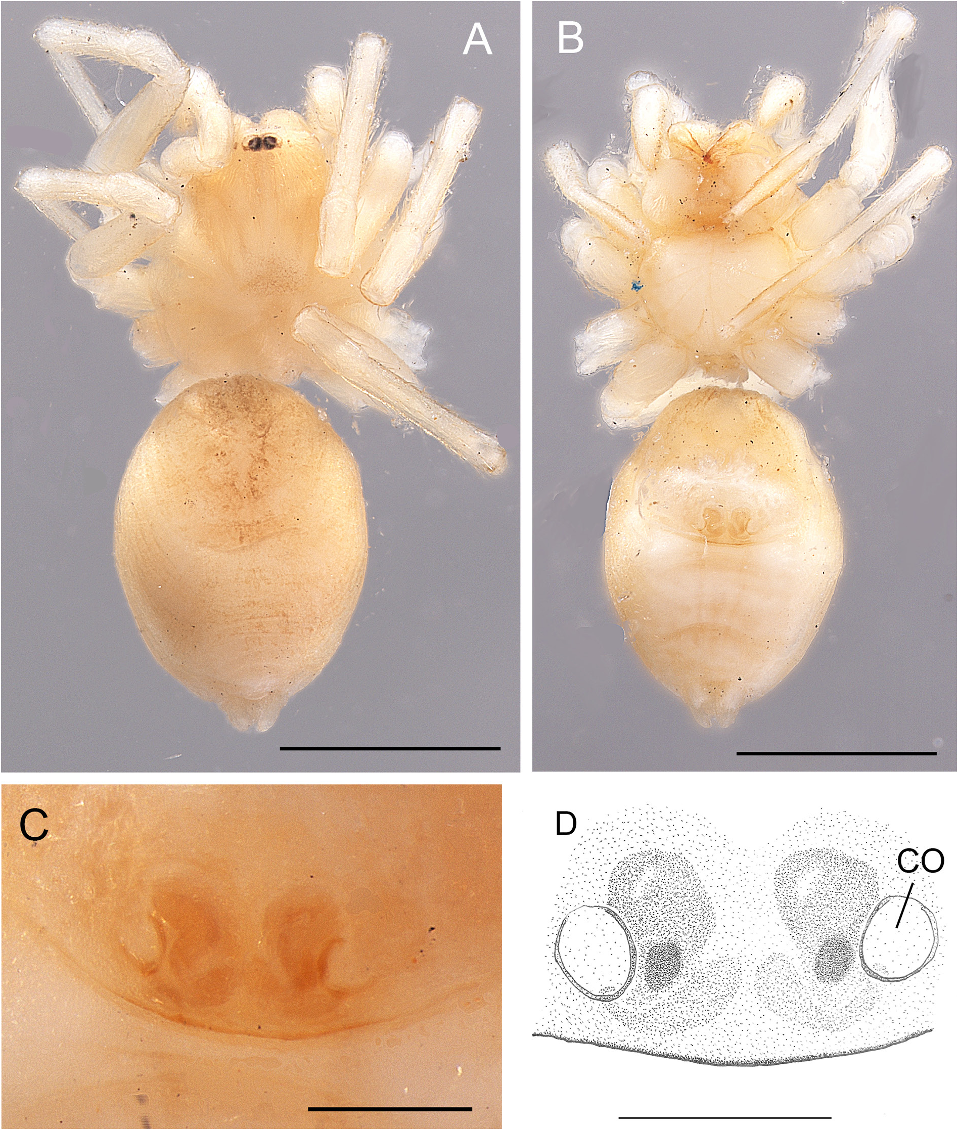
1. Sternum wider than long, no precoxal triangles, oval CO separated by three times their short axis, posterior eyes absent ( Fig. 5A, C–D View Fig ) .................................................... A. aethiopicum Simon, 1893 View in CoL
– Sternum not wider than long, with or without precoxal triangles ( Bosselaers & Jocqué 2002: fig 1k; Penniman 1985: 16), CO closer together, at least PLE present ( Figs 2B View Fig , 3 View Fig , 11D–E View Fig )........................ 2
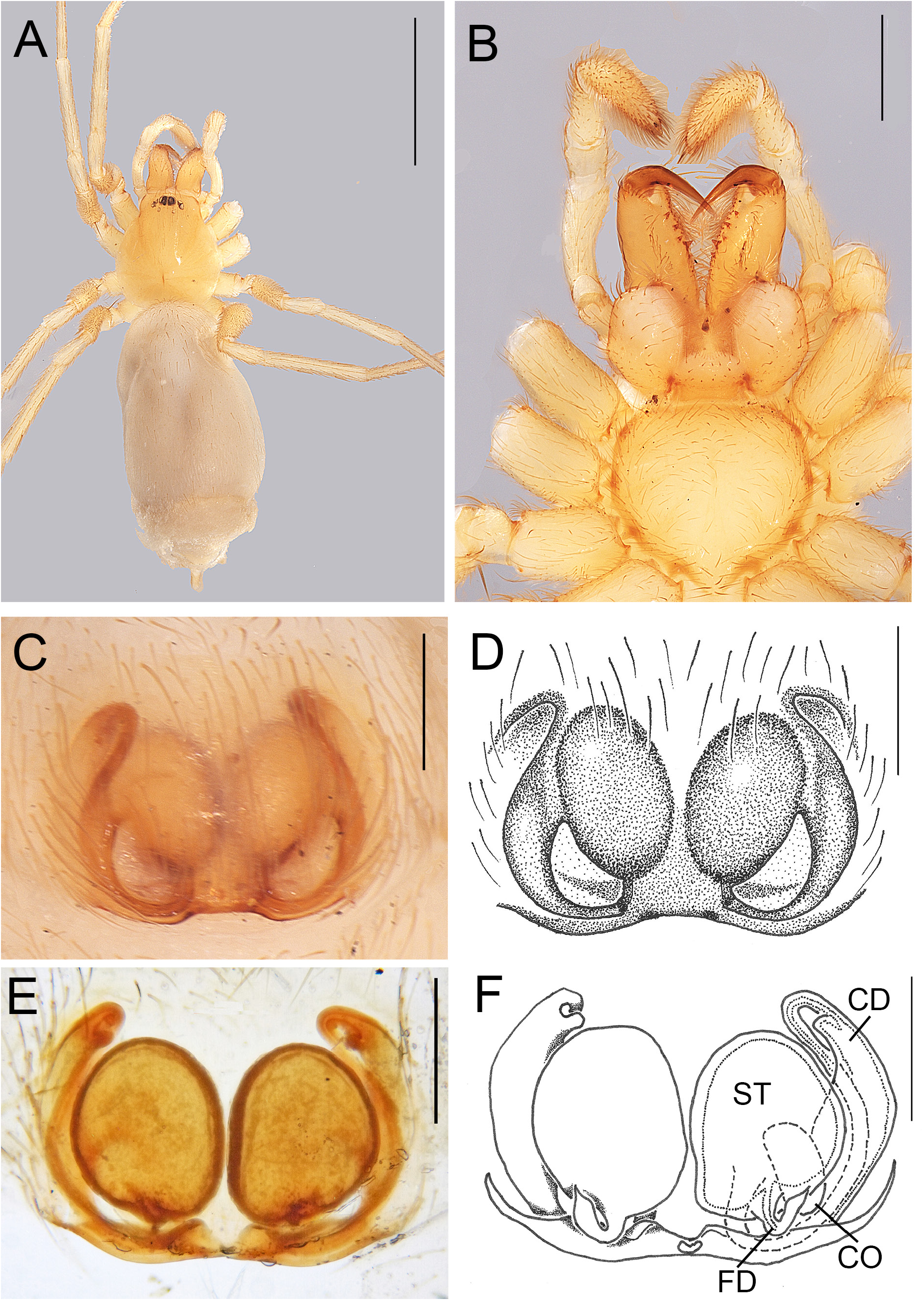
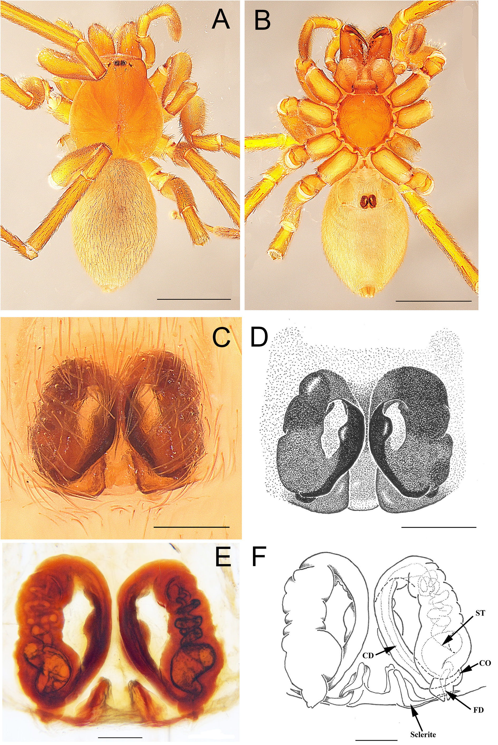
2. Sternum with strong precoxal triangles, epigyne more than twice as wide as long, CO transversely oval, separated by less than their long axis ( Fig. 11A, D–E View Fig ).......................... A. bouvieri Fage, 1936 View in CoL
– Precoxal triangles absent or epigyne not that wide or CO not transversely oval ( Fig. 33I–J View Fig ) .......... 3
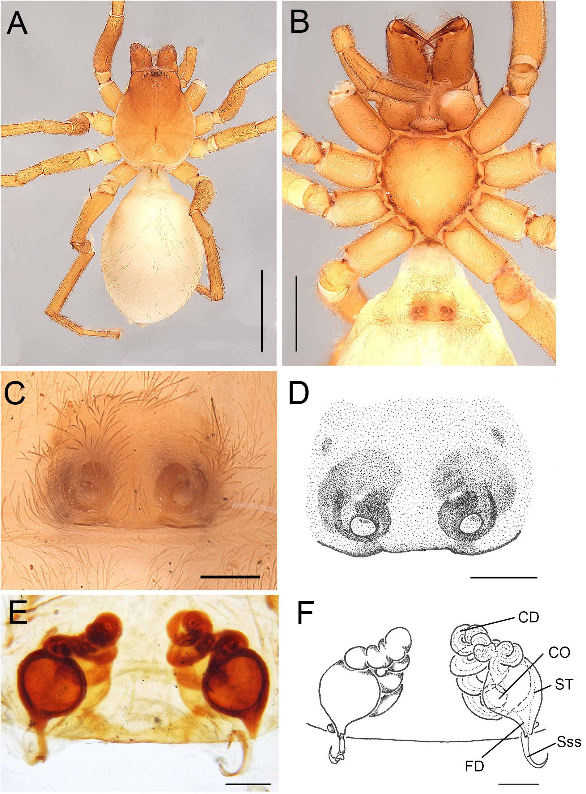
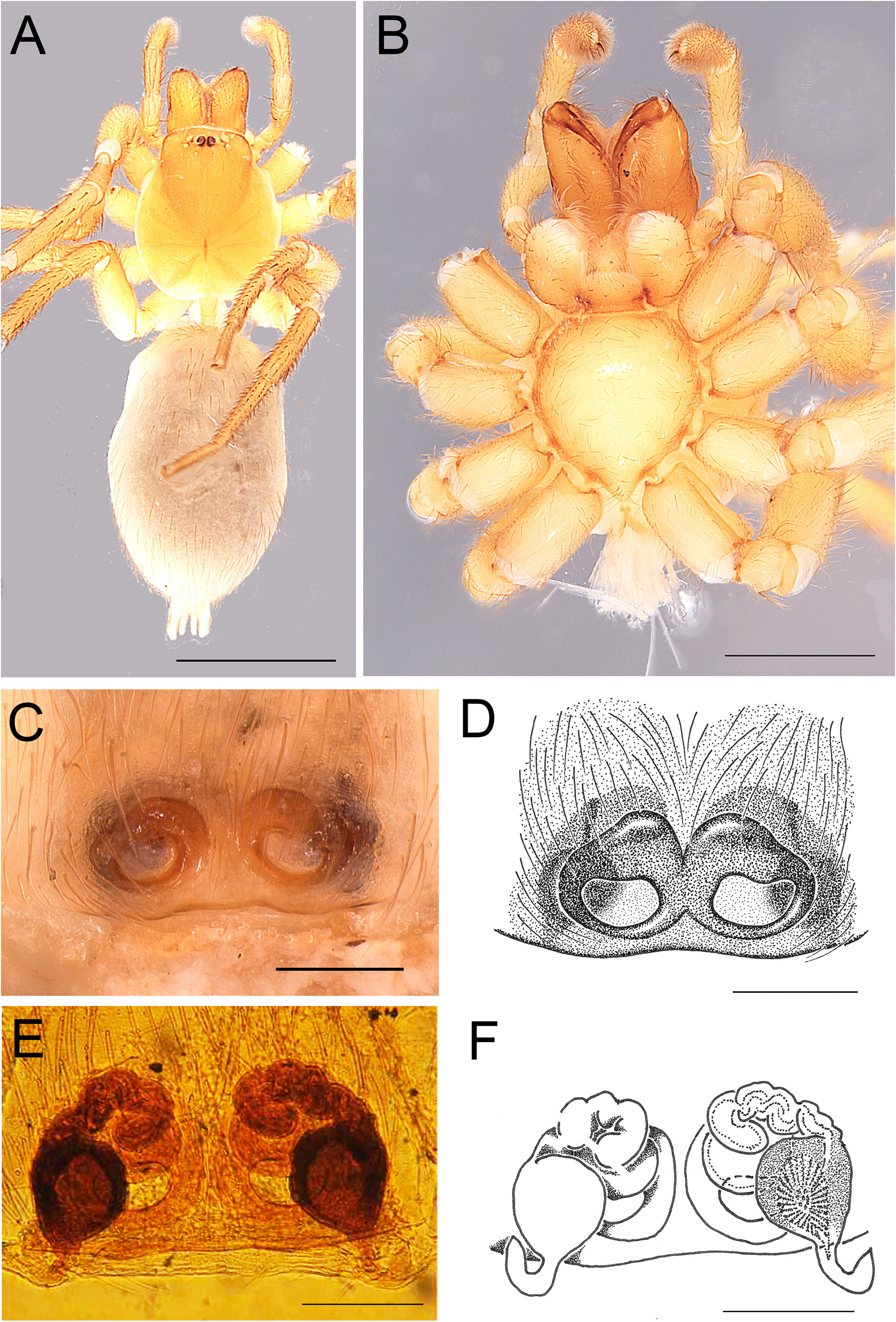
3. Copulatory openings longitudinally egg-shaped, separated by their short axis. Copulatory ducts winding, fused over their entire length into one dark brown, sclerotised mass ( Figs 33I–J View Fig , 35C–G View Fig ) ....................................................................................................................... A. raffrayi Simon, 1899 View in CoL
– Copulatory ducts not fused into one sclerotised mass over their entire length ................................. 4
4. Copulatory ducts simple, consisting of one to three mostly straight sections and showing one 180° bend (e.g., Figs 7C–D View Fig , 12E–F View Fig , 25E–F View Fig , 26E–F View Fig ) ................................................................................. 5
– Copulatory ducts narrower, long, helically coiled over at least part of their length (e.g., Figs 22E–F View Fig , 27E–F View Fig , 29E–F View Fig , 30E–F View Fig )..................................................................................................................... 12
5. Spermathecae very large, oval, medially located and almost touching, each connected to a laterally situated rather thin copulatory duct widening towards the CO ( Fig. 25E–F View Fig ). A. ghesquierei sp. nov.
– Spermathecae smaller, copulatory ducts wide, often with internal spikes, starting with a first, posterior stretch that runs transversely from the lateral side to the middle, followed by a second stretch running in anterior direction, a 180° bend and a third stretch running in posterior direction towards the spermathecae (e.g., Figs 23E–F View Fig , 26E–F View Fig )........................................................................ 6
6. Copulatory openings only vaguely defined ( Figs 7A–B View Fig , 26C–D View Fig )..................................................... 7
– Copulatory openings clearly defined, oval, kidney- or mung bean- ( Vigna radiata View in CoL ) shaped (e.g., Fig. 12B–D View Fig ) ....................................................................................................................................... 9
7. Animals small ( 3 mm), precoxal triangles weak or inconspicuous ( Fig. 16B View Fig ) ................................. 8
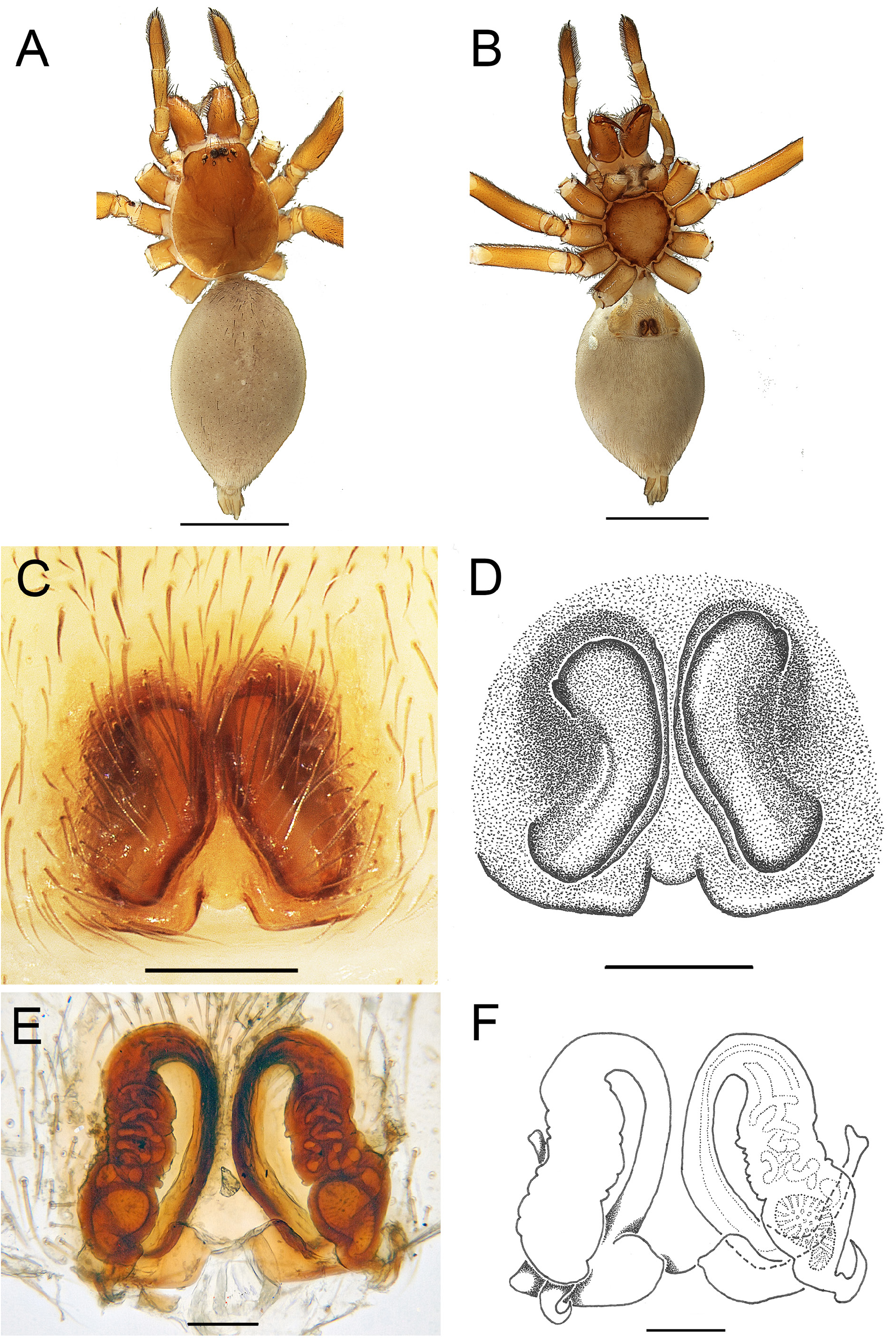
– Animals of medium size ( 6 mm), precoxal triangles pronounced ( Fig. 26B View Fig ), internal spikes of copulatory duct strong ( Fig. 26E–F View Fig ).................................................................... A. heligmos sp. nov.
8. Internal spikes of copulatory duct weak ( Fig. 7C–D View Fig )....................................... A. albinovani sp. nov.
– Internal spikes of copulatory duct strong ( Fig. 16E–F View Fig ).................................. A. delphiurum sp. nov.
9. Copulatory openings inversely comma-shaped ( Fig. 23C–D View Fig ) .................... A. elephantactes sp. nov.
– Copulatory openings oval or mung bean-shaped ( Fig. 12C–D View Fig ) ...................................................... 10
10. Copulatory openings transversely mung bean-shaped ( Fig. 12B–D View Fig ).................... A. cyamos sp. nov.
– Copulatory openings longitudinally circular or longitudinally egg-shaped ( Fig. 11C–D View Fig )...............11
11. Animals small ( 3 mm), sternum wider than long ( Fig. 9B View Fig )........................... A. anacardium sp. nov.
– Animals of medium size ( 5 mm), sternum as long as wide, third stretch of copulatory duct S-shaped, spermathecae narrow and tapering ( Fig. 18B, E–F View Fig )................................... ….. A. deogratias sp. nov.
12. Epigyne heavily sclerotised, consisting of two longitudinally oval or sausage-shaped plates. Copulatory openings longitudinally oval or inconspicuous ( Figs 22C–D View Fig , 37C–D View Fig )........................ 13
– Epigyne less heavily sclerotised, copulatory openings circular, transversely oval or transversely bean-shaped (e.g., Figs 29C–D View Fig , 30C–D View Fig )......................................................................................... 14
13. Epigynal sclerotised plates sausage-shaped, CO inconspicuous ( Fig. 22C–D View Fig )................................... ..................................................................................................................... A. divinagraciae sp. nov.
– Epigynal sclerotised plates ear-shaped, CO inconspicuous, situated in longitudinally oval depressions............................................................................................................... A. velum sp. nov.
14. Copulatory openings transversely kidney-shaped ( Figs 14C–D View Fig , 30C–D View Fig )....................................... 15
– Copulatory openings circular or transversely oval ( Figs 8C–D View Fig , 27C–D View Fig ) ....................................... 16
15. Second half of the copulatory duct with five or more closely appressed coils ( Fig. 14E–F View Fig ) .............. ................................................................................................................... A. cycnotrachelos sp. nov.
– Second half of copulatory duct with three more loosely appressed coils ( Fig. 30E–F View Fig ) ...................... ..................................................................................................................... A. ophiophagum sp. nov.
16. Copulatory openings small, oval, separated by three times long axis, first, anteriorly running stretch of copulatory duct consisting of four helical coils ( Fig. 29C–F View Fig ).................... A. katangensis sp. nov.
– Copulatory openings more closely spaced ( Figs 8C–D View Fig , 31C–D View Fig ).................................................... 17
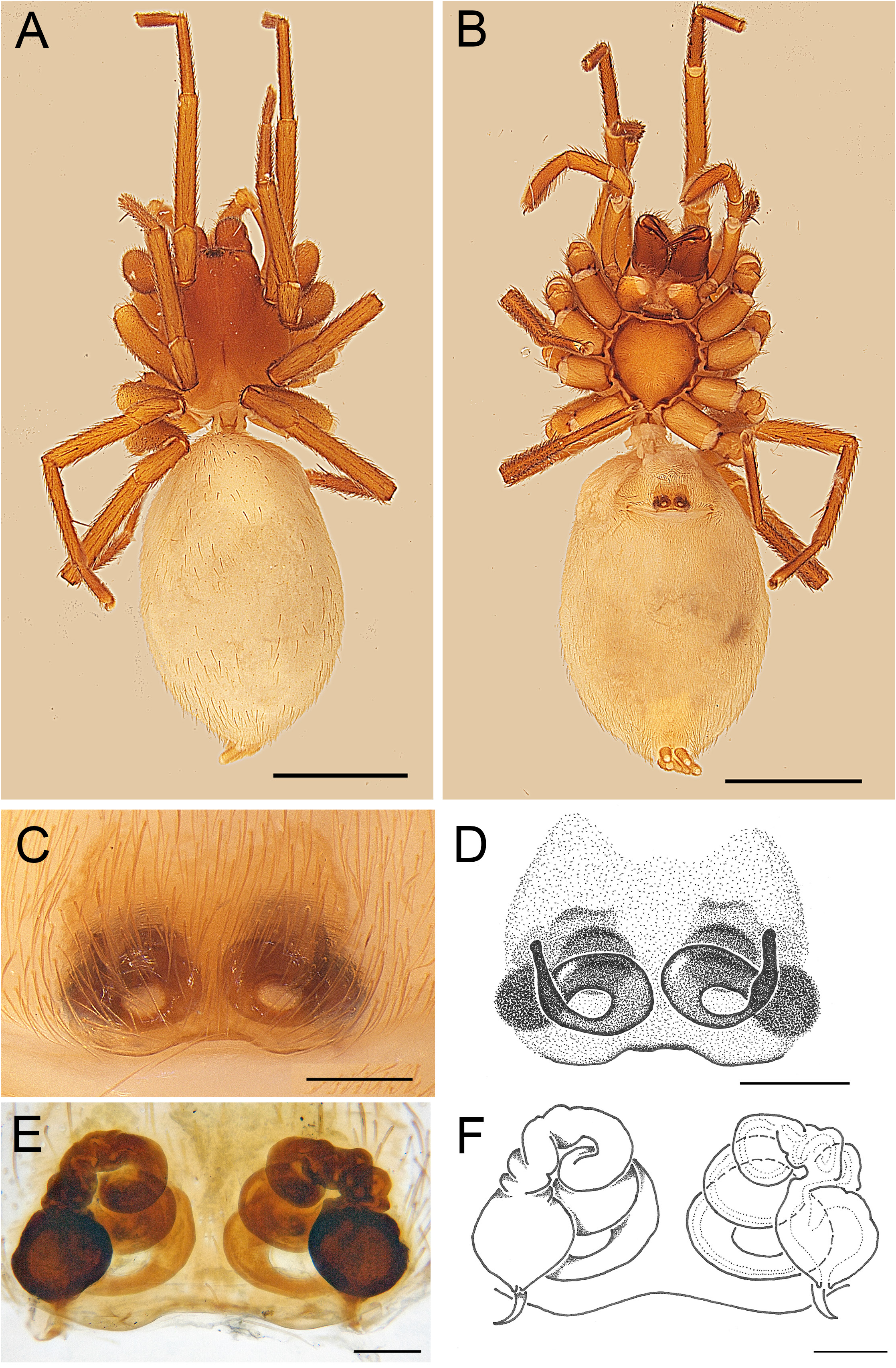
17. Animal small ( 3 mm), precoxal triangles weak, CO transversely oval, separated by two times their long axis, first stretch of copulatory duct consisting of five helical coils, spermathecae large, two thirds of vulva height ( Fig. 27B–F View Fig ) ........................................................................... A. helix sp. nov.
– Animals of medium size ( 5–7 mm), with strong precoxal triangles, first stretch of copulatory duct less extensively coiled, consisting of three helical coils ( Figs 8E–F View Fig , 31E–F View Fig ) ................................. 18
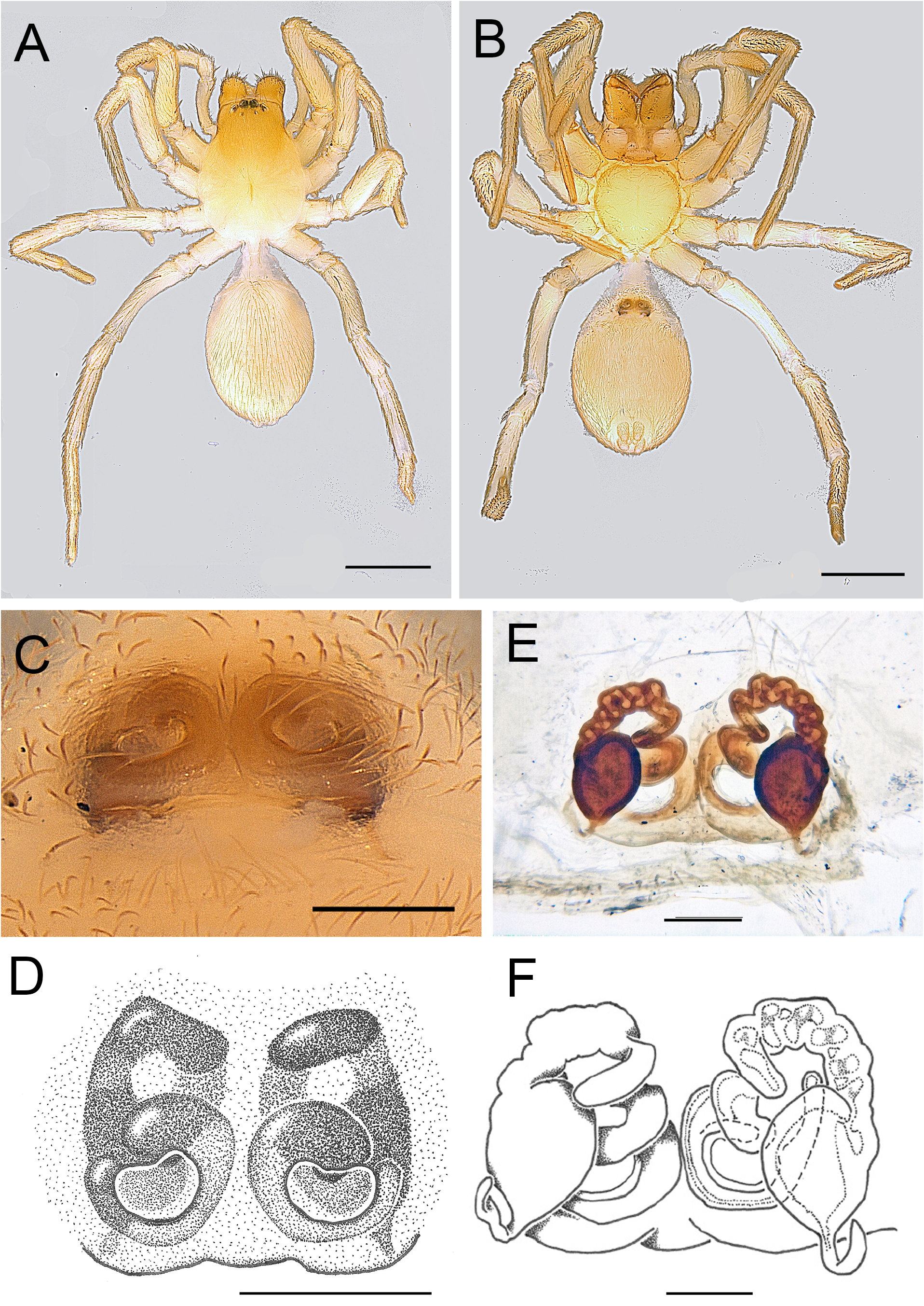
18. Epigyne consisting of two oval CO separated by two times their long axis and circled by a commashaped sclerotised ring ( Fig. 8C–F View Fig ).......................................................... …. A. alvoculatum sp. nov.
– Sclerotised part of epigyne one continuous plate, CO broadly oval and separated by their long axis ( Fig. 31C–F View Fig ) ...................................................................................................... A. prosopion sp. nov.


No known copyright restrictions apply. See Agosti, D., Egloff, W., 2009. Taxonomic information exchange and copyright: the Plazi approach. BMC Research Notes 2009, 2:53 for further explanation.