
Dilacreon ( Eluzalmon ) falcatus, Chen & Zhi, 2023
|
publication ID |
https://doi.org/10.11646/zootaxa.5347.1.1 |
|
publication LSID |
lsid:zoobank.org:pub:E9658506-5801-4B92-8140-A8FCE1EC8F40 |
|
DOI |
https://doi.org/10.5281/zenodo.8390882 |
|
persistent identifier |
https://treatment.plazi.org/id/91B06B52-E9D7-4FE2-B248-7A618BBB2BE4 |
|
taxon LSID |
lsid:zoobank.org:act:91B06B52-E9D7-4FE2-B248-7A618BBB2BE4 |
|
treatment provided by |
Plazi |
|
scientific name |
Dilacreon ( Eluzalmon ) falcatus |
| status |
sp. nov. |
Dilacreon ( Eluzalmon) falcatus sp. nov.
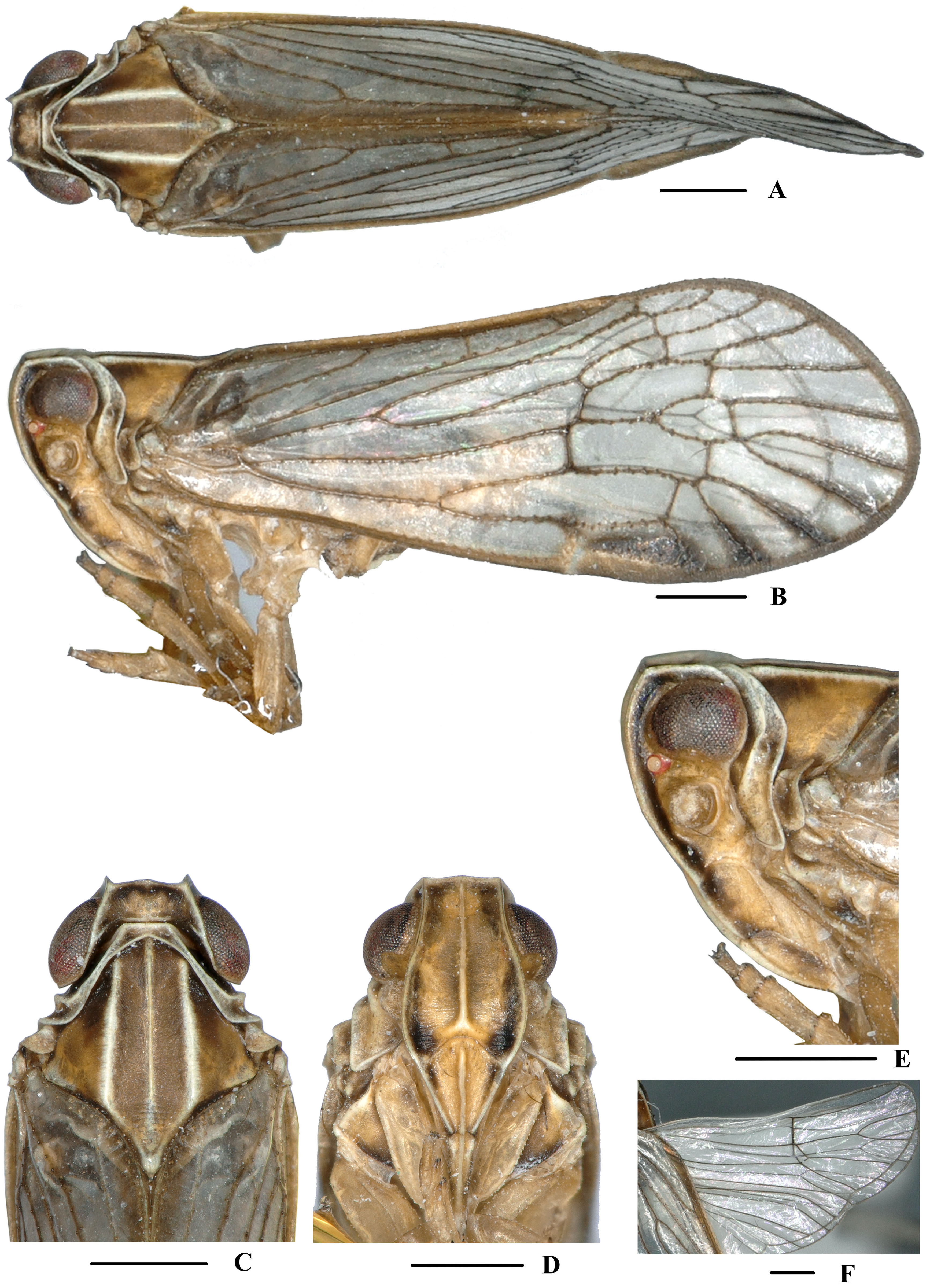
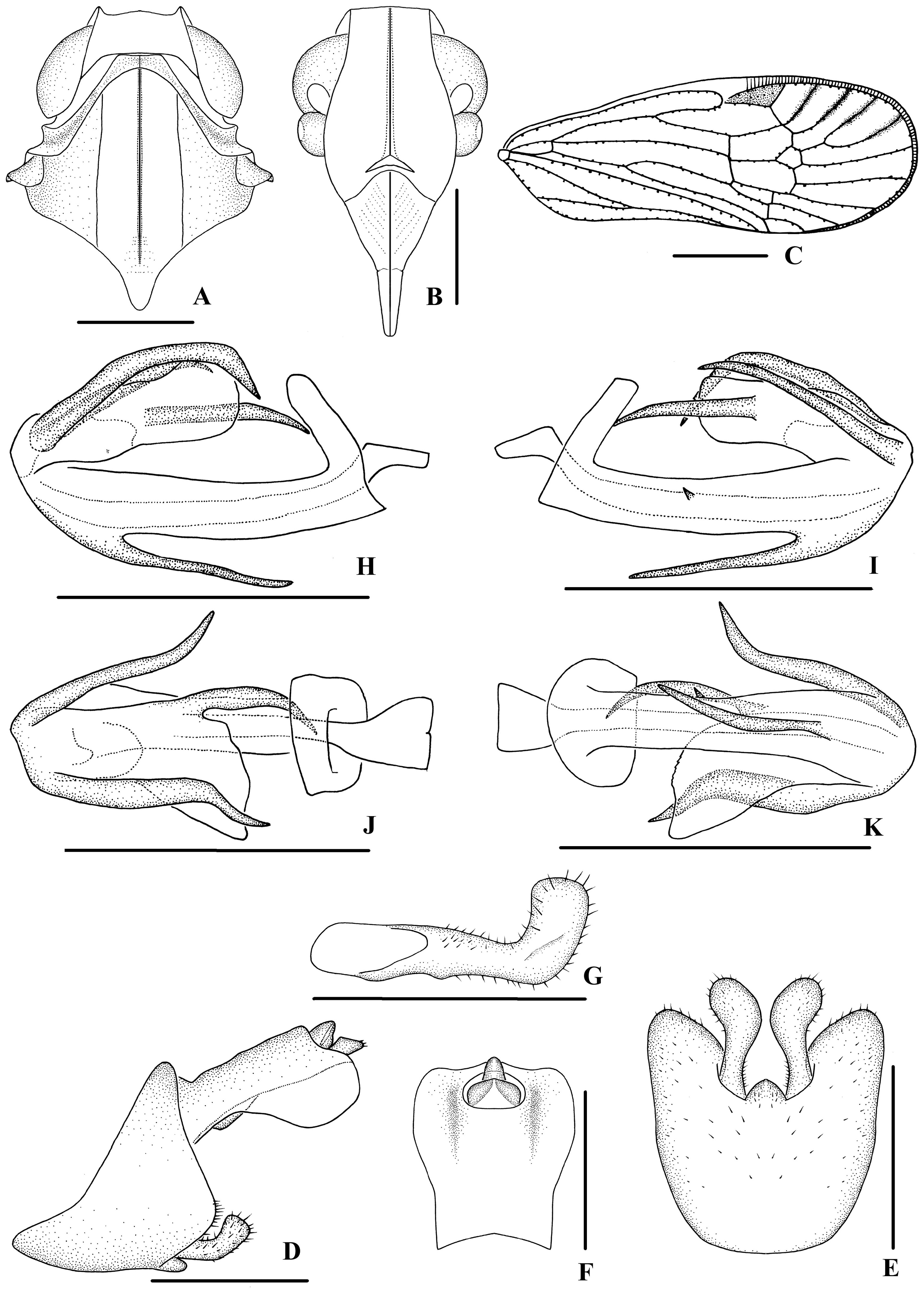
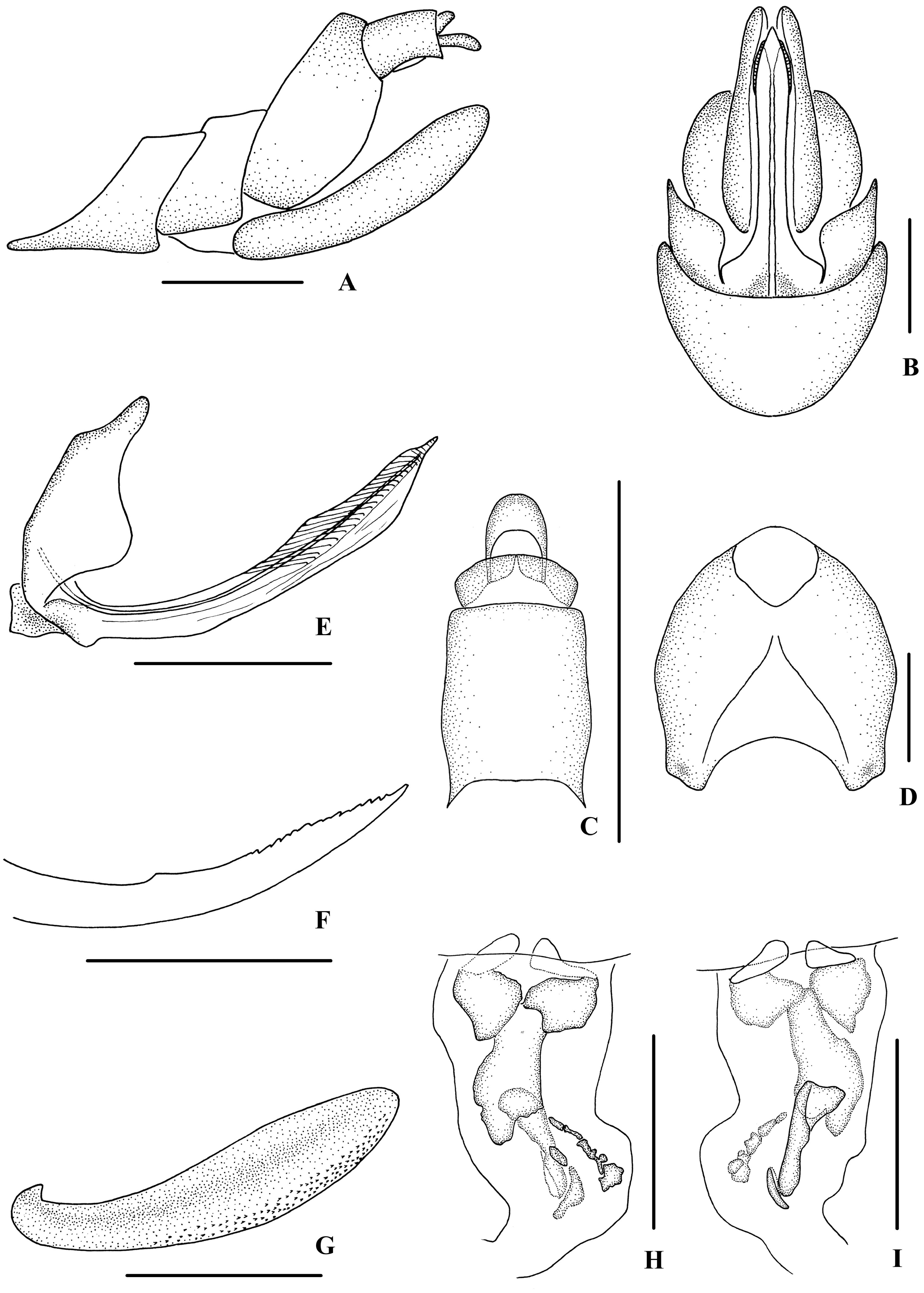
( Figs 19‒21 View FIGURE 19 View FIGURE 20 View FIGURE 21 )
Description. Body length: male 5.1‒5.4 mm ( n = 5), female 6.1‒6.5 mm ( n = 8).
Coloration. General color grayish brown ( Fig. 19A–F View FIGURE 19 ). Eyes blackish brown, ocelli light yellow, semitransparent. Vertex generally brown with lateral carinae yellowish brown. Face generally yellowish brown with lateral margins dark brown; rostrum yellowish brown. Pronotum and mesonotum brown with light yellow carinae. Forewing grayish brown, semi-translucent, veins darker; stigma dark brown. Hind tibiae and abdominal sternites yellowish brown.
Head and thorax. Vertex ( Figs 19C View FIGURE 19 , 20A View FIGURE 20 ) broad, 2.7 times wider than long; anterior margin slightly convex, median carina obsolete, posterior margin archedly recessed. Frons ( Figs 19D View FIGURE 19 , 20B View FIGURE 20 ) widest slightly below the level of antennae, 1.3 times as wide as long; frontoclypeal suture nearly concave into an arch; middle carina complete; lateral carinae distinct and slightly elevated. Pronotum ( Figs 19C View FIGURE 19 , 20A View FIGURE 20 ) 2.4 times longer than vertex; median carina indistinct, posterior margin nearly at right angle. Mesonotum 1.7 times longer than pronotum and vertex combined. Forewing ( Fig. 20C View FIGURE 20 ) 2.7 times longer than wide, with 10 apical and 6 subapical cells; fork Sc+RP distad of fork CuA 1 +CuA 2, first crossvein r-m slightly basad of fork MP, RP two branches, MP with five terminals: MP 11, MP 12, MP 2, MP 3, and MP 4, fork MP 1 +MP 2 basad of fork MP 3 +MP 4. Metatibiotarsal formula: 6/7/8, second segment of hind tarsus with three platellae.
Male genitalia. Pygofer ( Fig. 20D, E View FIGURE 20 ) symmetrical, dorsal margin concave and U-shaped, slightly widened towards apex in ventral view; in lateral view, lateral lobes triangularly extended caudally, medioventral process triangular in ventral view. Anal segment ( Fig. 20D, F View FIGURE 20 ) broad, dorsal margin almost straight, apical half of ventral margin convex, apical lobes round in lateral view; 1.2 times longer than wide in dorsal view; anal style strap-shaped, slightly beyond anal segment. Gonostyli ( Fig. 20D, E, G View FIGURE 20 ) symmetrical in ventral view; in inner lateral view, base of ventral margin slightly concave, dorsal margin bending inwards in a right-angle arc in the middle, apical part slightly extended, apical margin round. Aedeagus ( Fig. 20H–K View FIGURE 20 ) with total of five processes. The ventral margin of periandrium with a slender spinous process near apex, relatively straight, apex ventrocephalically directed; left side in the middle with a very short spinous process, dorsocephalically directed. Endosoma (=flagellum) moderately sclerotised, medium-sized, generally dorsally curved. Base of right side with a long and broad falcate spinous process, which basal half pointed upward and then right-ventrocephalically directed. Left side with two spinous processes, one originated from base, left-cephalically directed, the other one on the apex, medium-sized, slightly curved and right-ventrocephalically directed.
Female genitalia. Posterior margin of pregenital sternite concave. Tergite IX ( Fig. 21A, D View FIGURE 21 ) moderately sclerotised, 1.2 times longer than wide in caudal view. Anal tube ( Fig. 21A, C View FIGURE 21 ) short, nearly rectangular, 1.4 times longer than wide in dorsal view; dorsal margin nearly straight and ventral margin slightly concave in lateral view, anal styles strap-shaped. Gonapophysis VIII ( Fig. 21E View FIGURE 21 ) elongate, and slightly curved upwards. Gonapophysis IX ( Fig. 21F View FIGURE 21 ) with one middle tooth, at a distance ratio, between middle tooth to apex and length of denticulate portion, of 1.8. Gonoplac ( Fig. 21G View FIGURE 21 ) rod-like, 4.0 times longer than wide. Posterior vagina ( Fig. 21H, I View FIGURE 21 ) elongate. The ventral wall of posterior vagina with three large sclerites, two nearly round at base and one long longitudinal sclerite in the middle aera; several small sclerites on the right distal half, elliptic or irregular in shape, arranged in an oblique row, and a nearly oval sclerite located on the left side of the queue. The dorsal wall with a long longitudinal large sclerite and a small elliptic sclerite on distal half.
Type material. Holotype: ♂, CHINA: Bakaxiaozhai ( 21°58’N, 101°13’E), Mengla County, Yunnan Province, 15 June 2016, leg. Ying-Jian Wang and Qiang Luo; GoogleMaps paratypes: 3♂♂ 3♀♀, same data as holotye; GoogleMaps 1♂ 5♀♀, Mohan Town, Mengla County , Yunnan Province, 20‒22 June 2016, leg. Ying-Jian Wang, Qiang Luo and Liang-Jing Yang. GoogleMaps
Host plant. Unknown.
Distribution. China ( Yunnan).
Remarks. Male genitalia of D. ( E.) falcatus sp. nov. are similar to those of D. ( E.) chionomus Fennah, 1980 , but differ in: (1) the ventral margin of periandrium with a spinous process (the latter without spinous process in the same position); (2) left side of periandrium at middle with a very short spinous process (the latter with one spinous process on dorsal margin and left side); (3) right side of endosoma with one spinous process (right side of endosoma with two spinous processes in D. ( E.) chionomus ).
Etymology. The specific name refers to apex of right side of endosoma with a long and broad falcate spinous process.
No known copyright restrictions apply. See Agosti, D., Egloff, W., 2009. Taxonomic information exchange and copyright: the Plazi approach. BMC Research Notes 2009, 2:53 for further explanation.