
Neoperla spio (Newman, 1839) Dr.
|
publication ID |
https://doi.org/10.11646/zootaxa.5316.1.1 |
|
publication LSID |
lsid:zoobank.org:pub:BC922E16-2614-4F3D-AD82-87A845DE7E2B |
|
DOI |
https://doi.org/10.5281/zenodo.16763865 |
|
persistent identifier |
https://treatment.plazi.org/id/E12C876C-4A3C-FFDC-FF4F-FE46FA0C091A |
|
treatment provided by |
Plazi |
|
scientific name |
Neoperla spio |
| status |
|
IV.3. The Neoperla spio View in CoL -complex (~ clade G)
In males, a pale line of weak sclerotisation runs across the middle of T7 which caudomedially has a pyramid-shaped process, with SB near tip and along the sclerotised underside. Median sclerite on T8 raised, with a hump or a process, the penis is tubular and sclerotised.
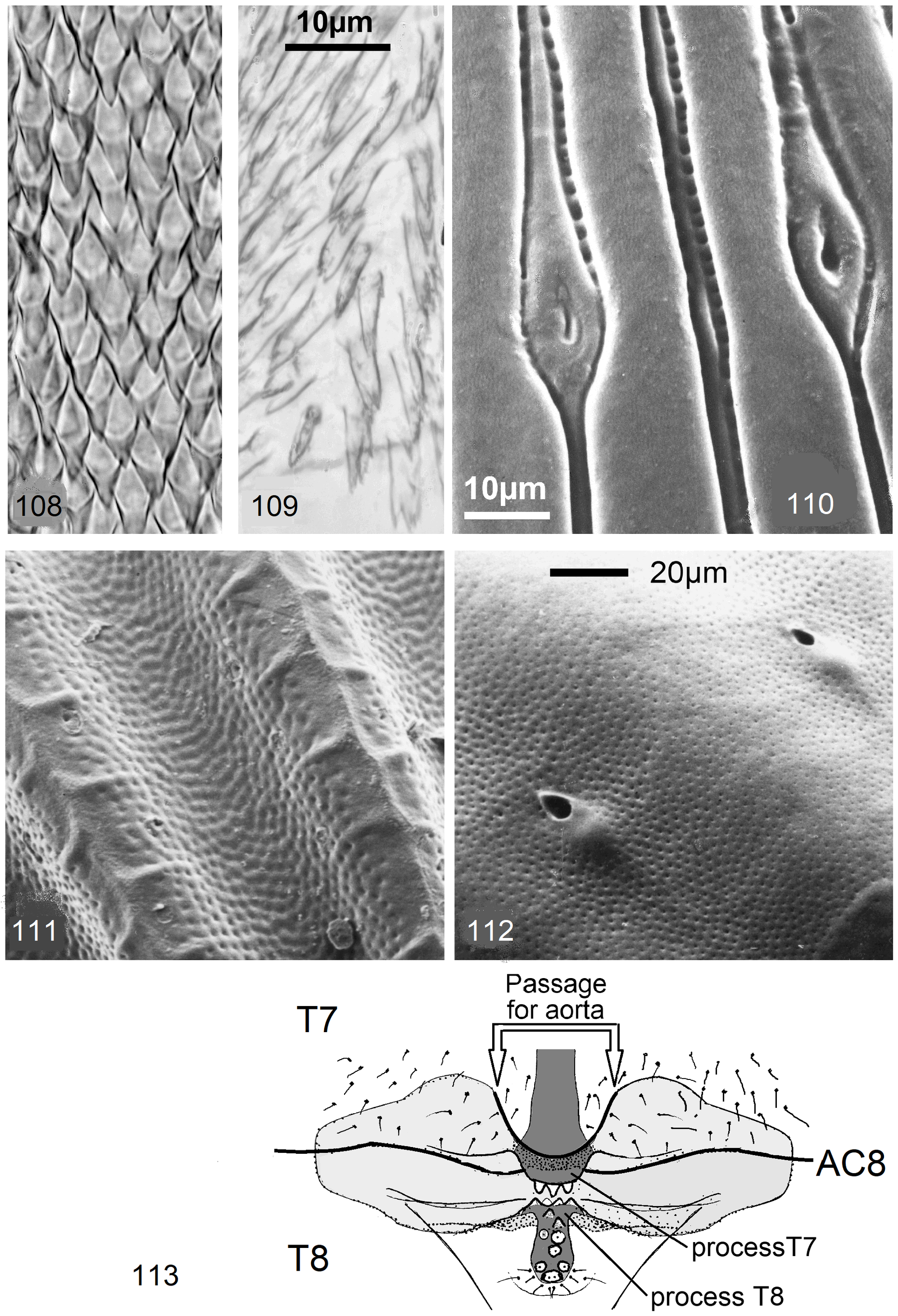
In several males, especially in the N. burgeoni -complex where males can raise a median sclerite on T8, the antecosta of T8 has paramedian apodemes projecting into segments 7 and 8, between the apodemes remains a passage for the aorta ( Fig. 113 View FIGURES 108–113 ). The structure acts like a hinge, longitudinal muscles from antecosta 7 and attached to the apodemes affect both T7 and T8: the ventro-median ridge of the process on T7 is exposed and directed backward, resembling a vertical keel on the caudal edge of T7. Comparable modifications of the antecosta 8 occur also in some other species but are less strongly developed.
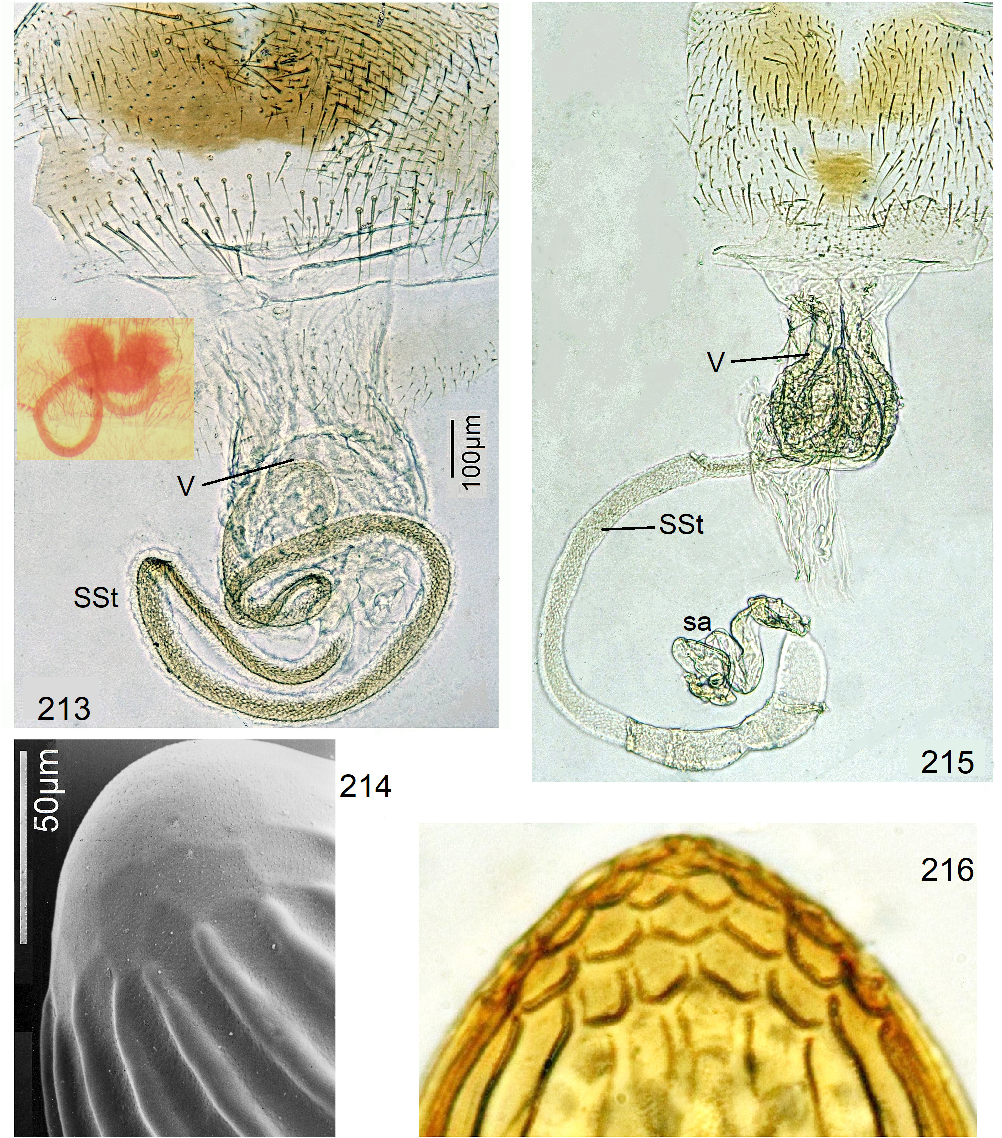
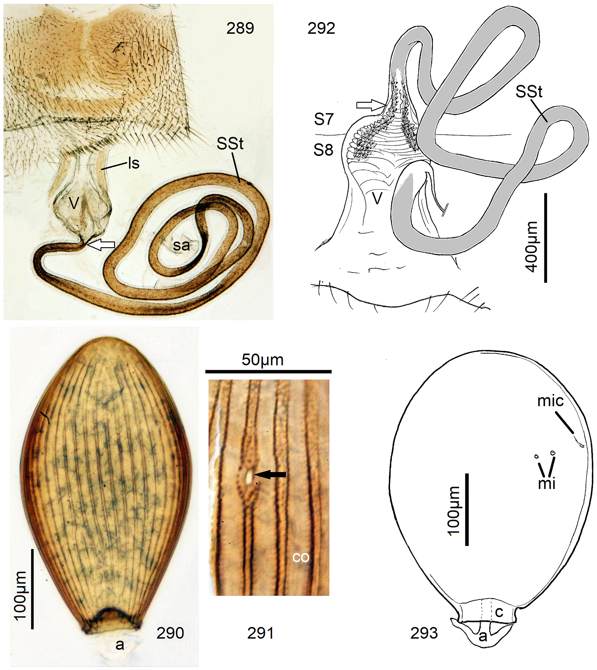
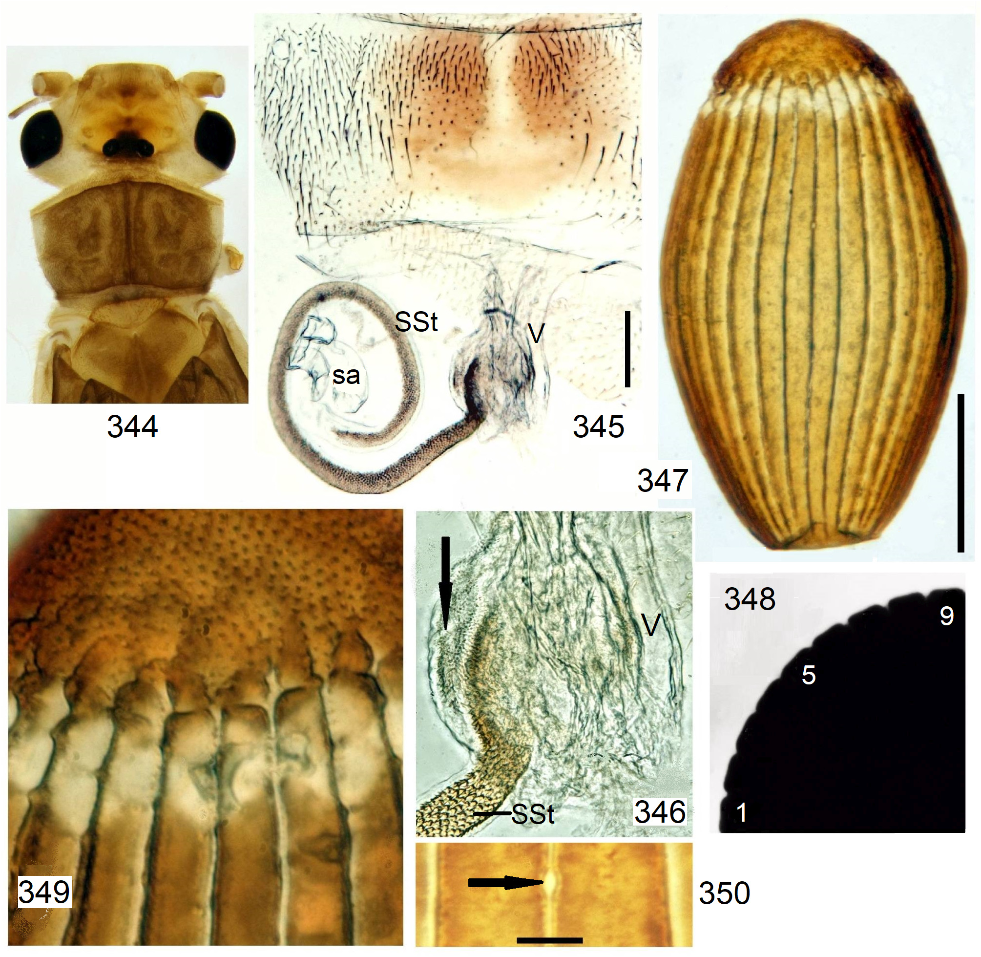
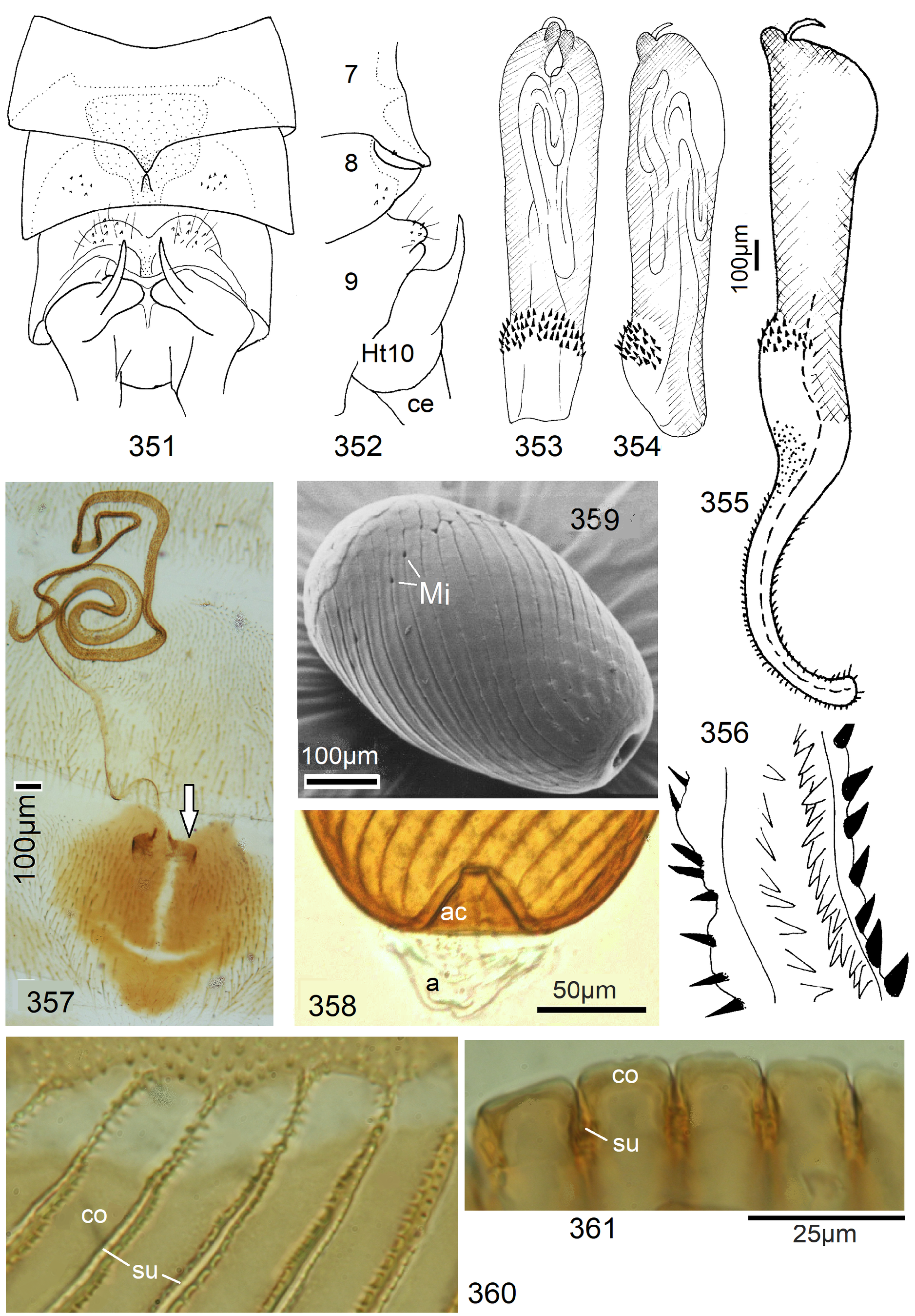
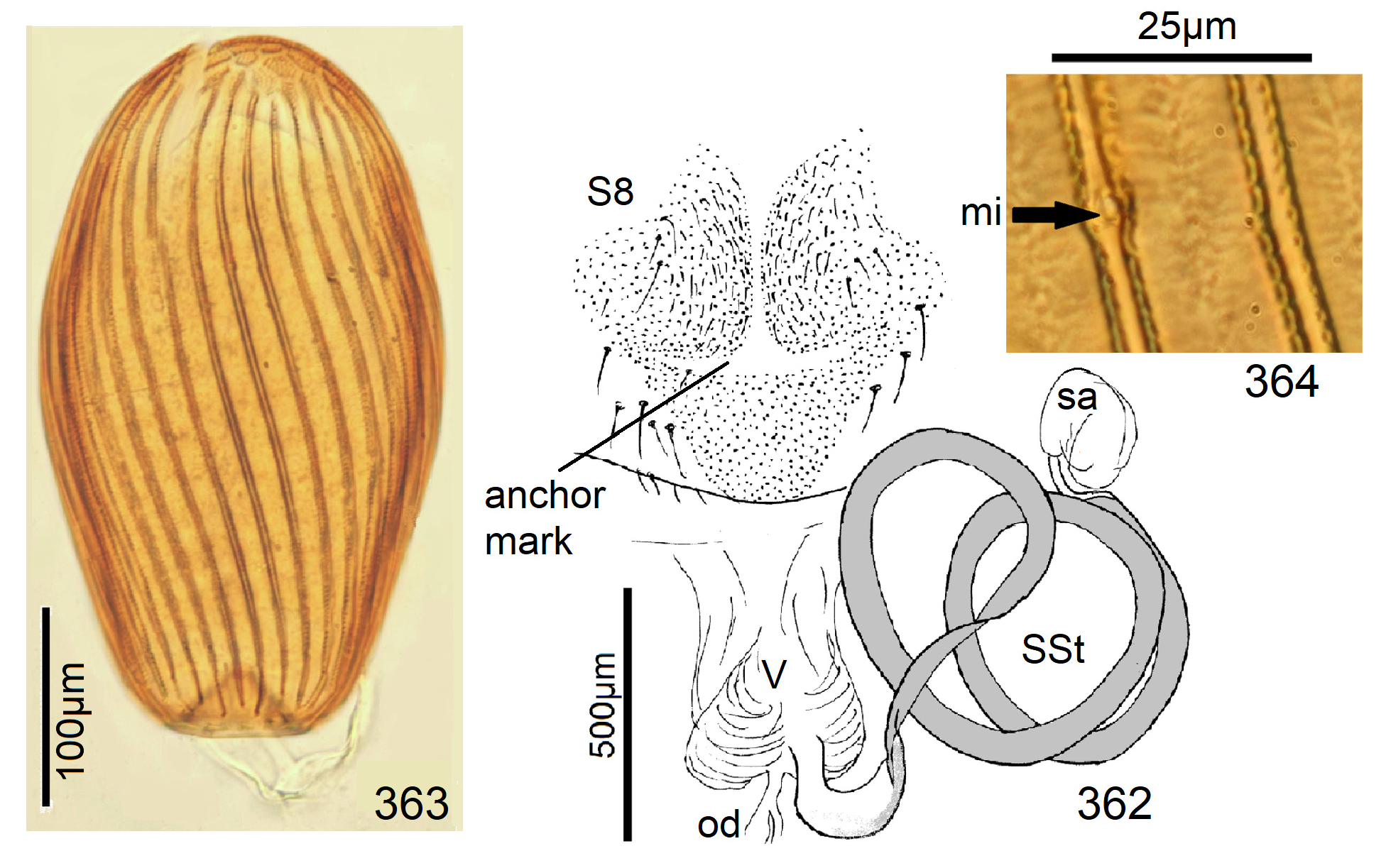
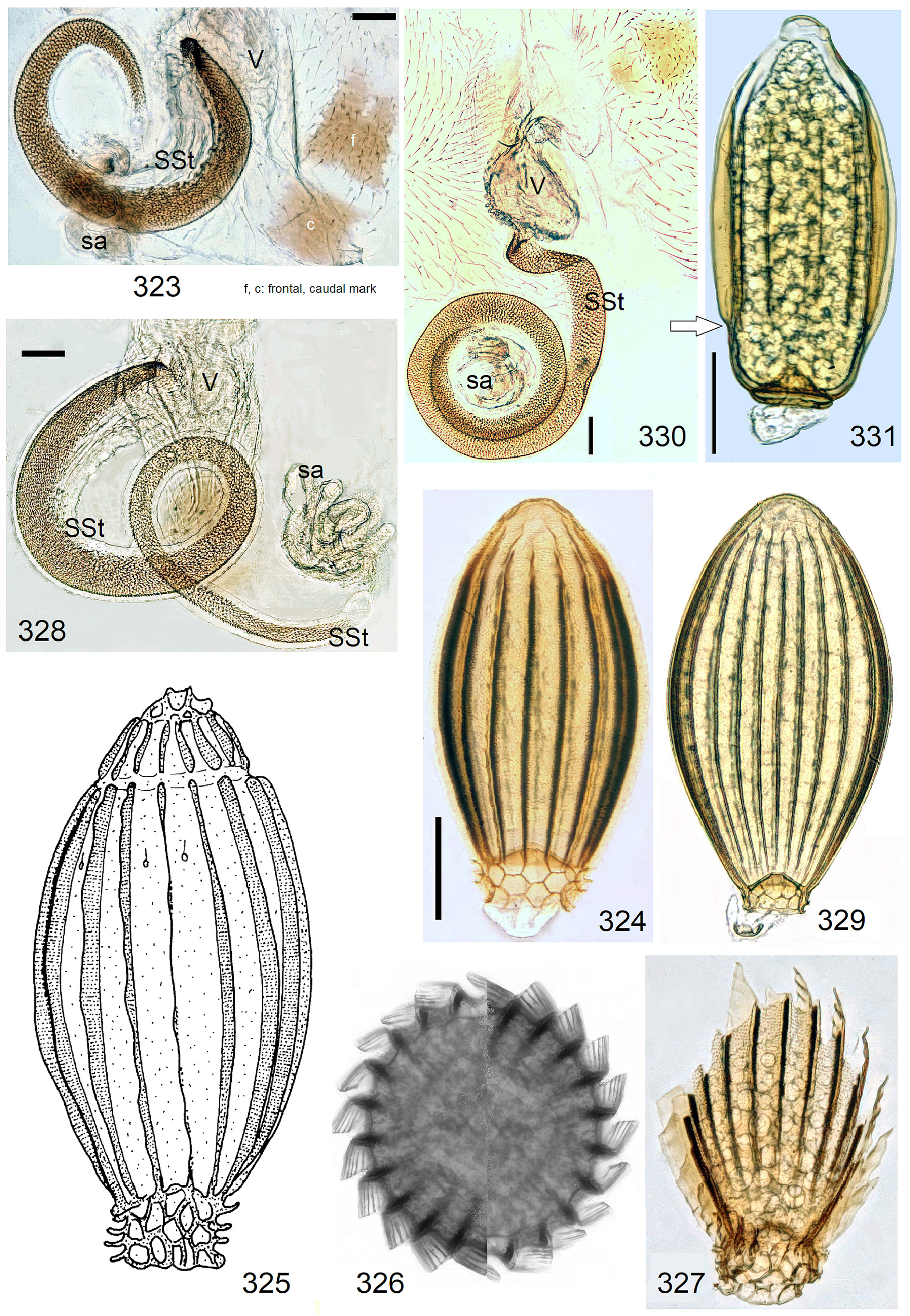
Female S8 with unmodified caudal edge, well pigmented females with three brown sclerites forming a triangle with caudal tip (e.g., Fig. 215). In variants of this pattern, sclerites may be large and merge around a pale center resembling an anchor (e.g., Fig. 264 View FIGURES 258–266 ). In several species the caudal sclerite is weak or absent and only the strong paired anterior sclerites remain (e.g., Fig. 213 View FIGURES 213–214 ). Anchor patterns on S8 occur also in other species groups (e.g., Figs. 289 View FIGURES 289–293 , 345 View FIGURES 344–350 , 357 View FIGURES 351–361 , 362 View FIGURES 362–364 ). Females of the N. dubia - and the N. orthonema -complexes differ and have on S8 2 brown marks in a longitudinal row which may be difficult to see in pale specimens ( Figs. 323, 330 View FIGURES 323–331 ).
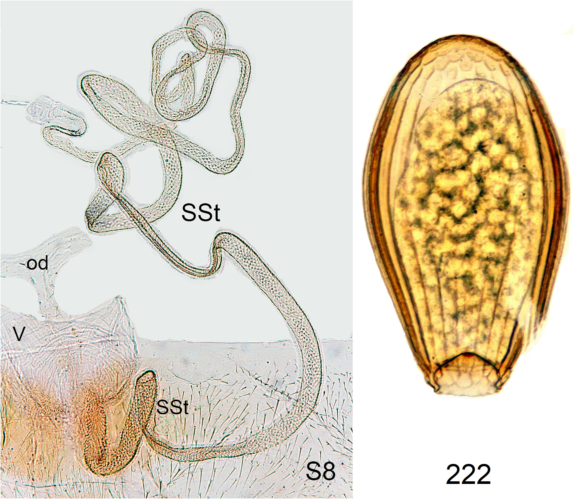
The vagina is sometimes with spines laterally from the SSt-attachment, otherwise unmodified. The SSt is coiled and of variable length, its opening into the vagina is narrower than in the transvaalensis -group (e.g., Figs 226 View FIGURES 223–226 , 236 View FIGURES 231–237 , 244 View FIGURES 244–249 ). The narrow concave edge of the coil lacks scales and is membranous. The bare edge widens next to the vagina, the length of the basal section varies, sometimes in contains longitudinal folds and seems to be extensible. The rest of the SSt has a dense cover of brown scales resembling roof tiles or fish scales ( Fig. 108 View FIGURES 108–113 ).
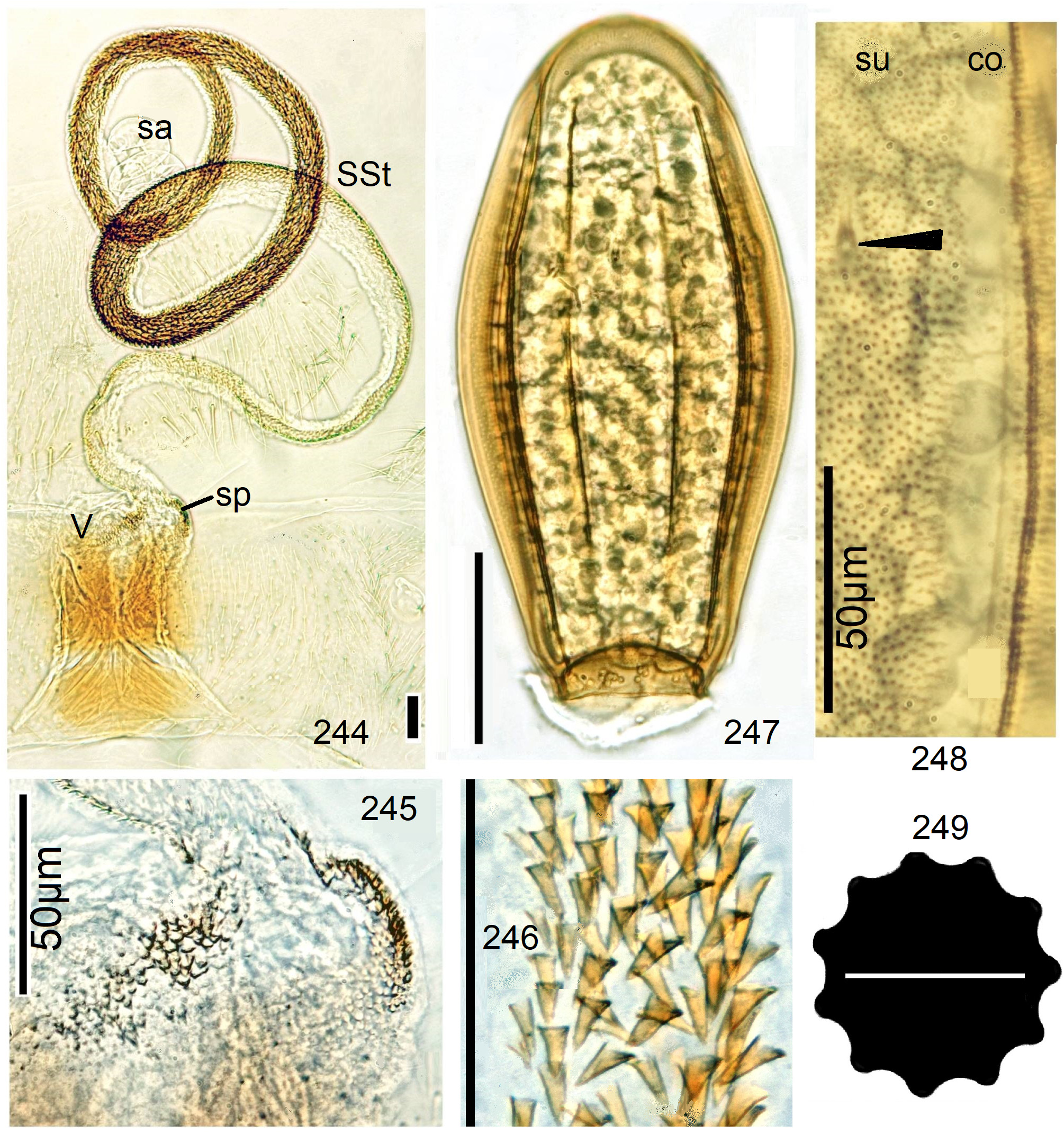
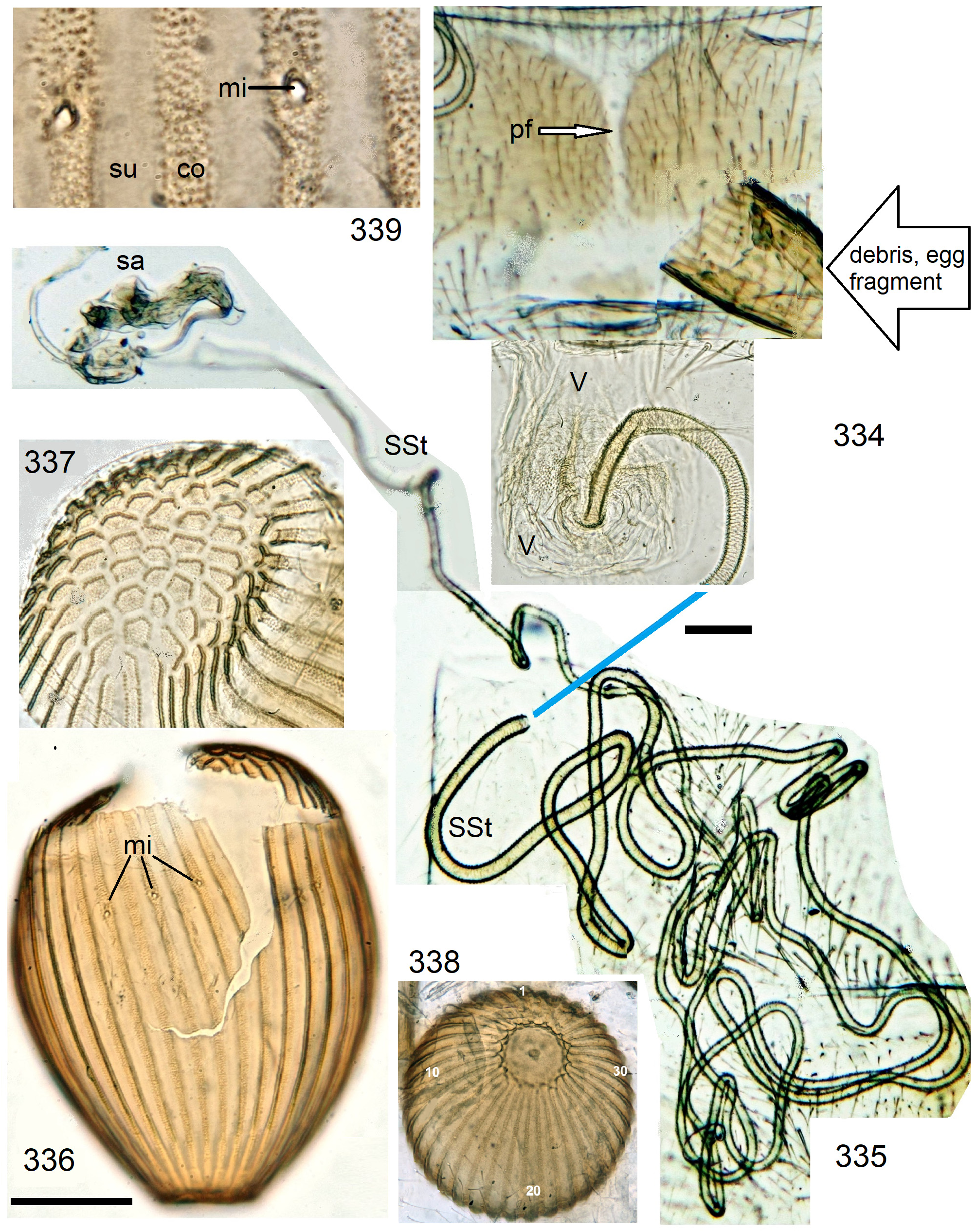
Eggs have straight striae, sulci are about as wide as the impunctate costae (e.g., Figs. 214 View FIGURES 213–214 , 339 View FIGURES 334–339 ), punctation in the sulci without order, micropyles freely visible (e.g., Fig. 257 View FIGURES 250–257 ). Most species have 25– 30 egg striae, but N. spio has only about a dozen ( Fig. 247 View FIGURES 244–249 ).
The analyses of DNA sequence data support strongly (all-NT: 65.7/100/90) to very strongly (mt-NT) the monophyly of the N. spio -complex + N. orthonema- complex (clade G, including clade H; see Figs. 491–492 View FIGURE 491 View FIGURE 492 , 496 View FIGURE 496 ).
Keys to species in the N. spio View in CoL -complex
The spio View in CoL -group was originally defined by morphology. The key covers all originally included species, regardless of whether membership in the group was subsequently confirmed by DNA (clades G + H) or not.
Males (unknown of N. gordius , N. spaghetti , N. sassandrae , N. benti )
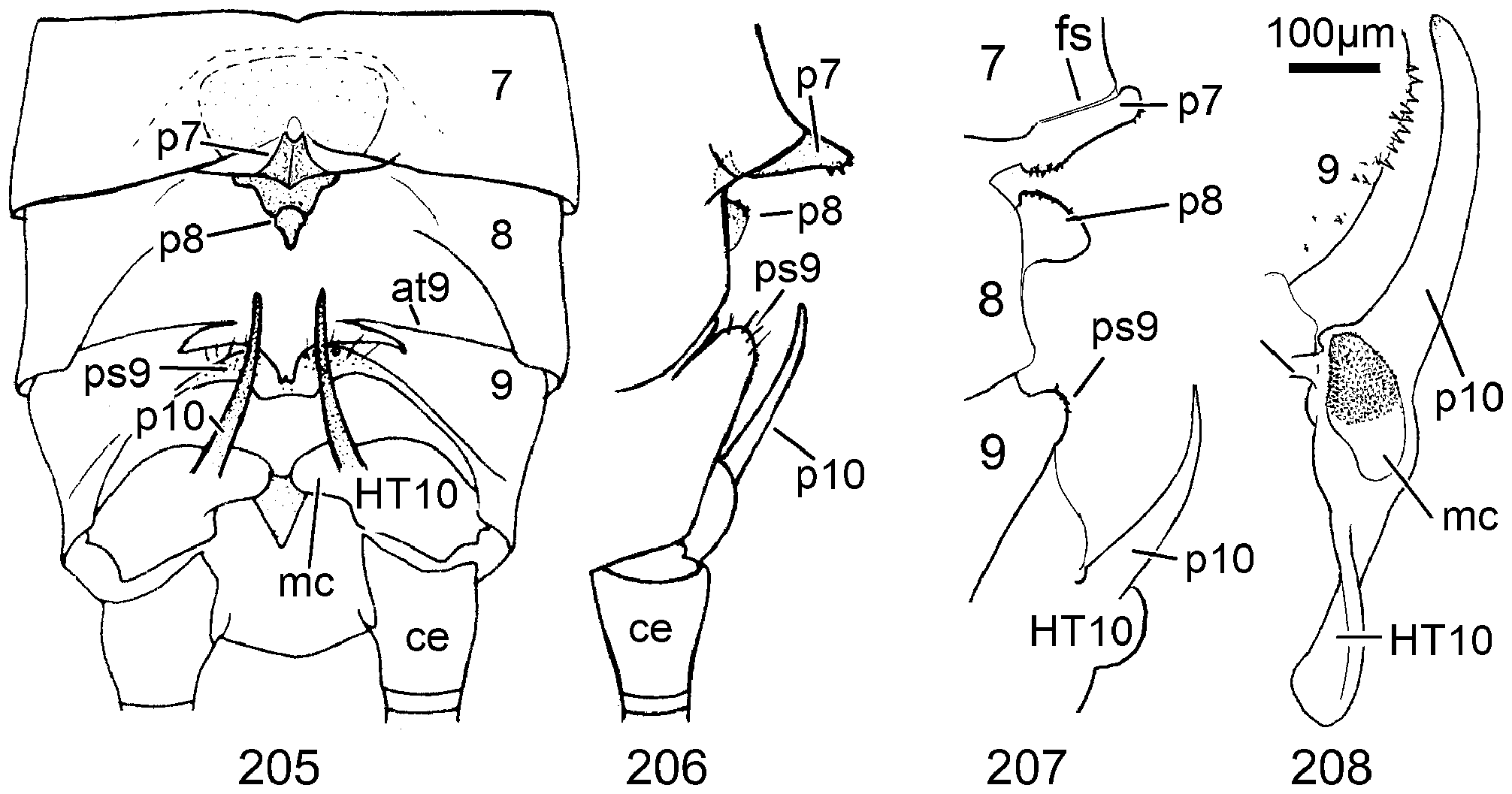
1 T8 with a longitudinal sclerite that can be raised. In lateral view the raised sclerite resembles a kidney. A fine pale suture surrounds the button-like top of the T7 process in front. HT10 long, slender, regularly arched ( Figs. 205–208 View FIGURES 205–208 )............ 2
1' T8 without movable sclerite or process, remainder variable. Species with narrow tube-like endophallus belong here....... 4
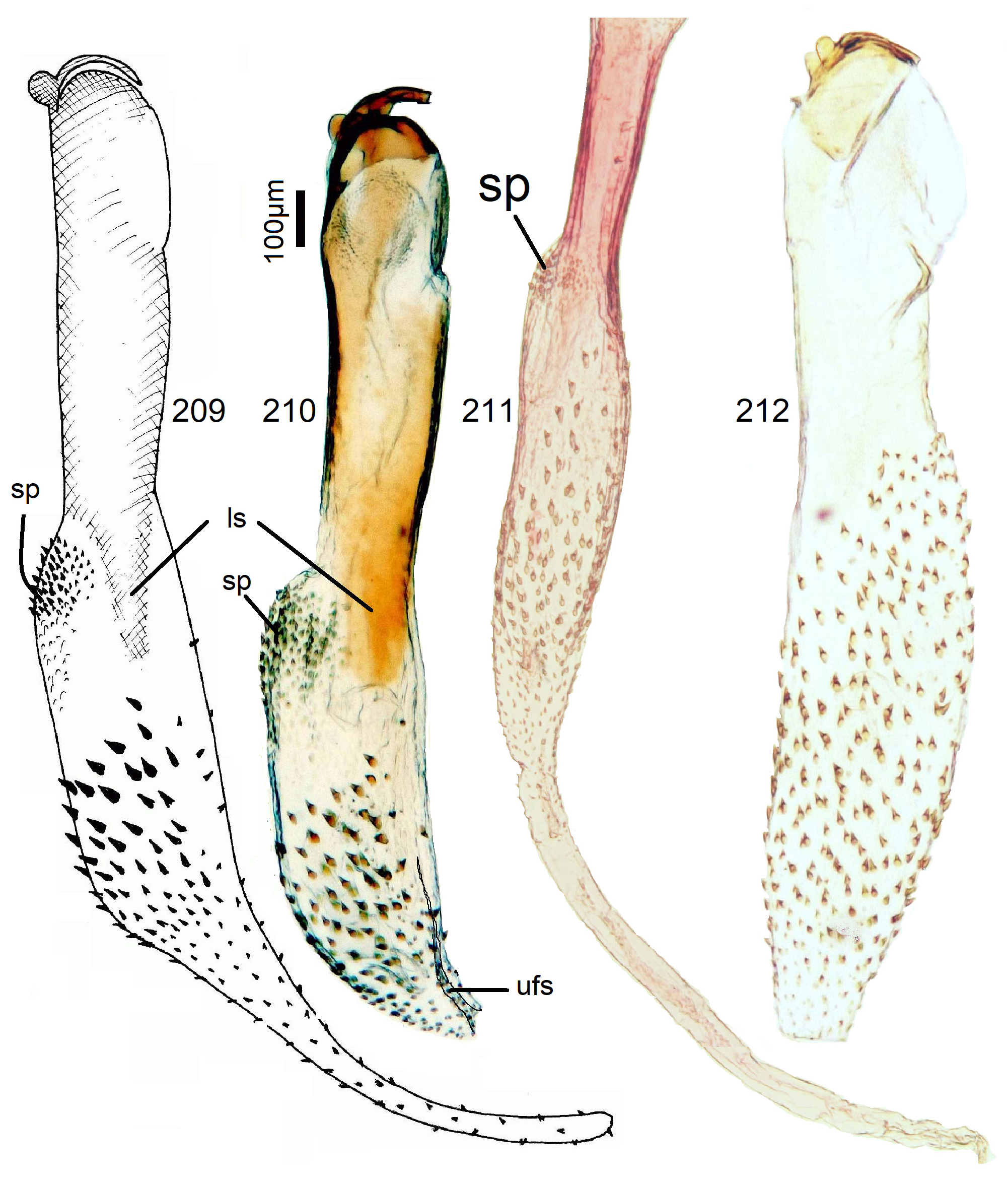
2 Penis apex with elongate lateral sclerites between membranous ventral and dorsal areas and with a transverse dorsodistal patch of spines. Endophallus much wider than the tube, base bare. The spiny section shows a size gradient from large dorsobasal to smaller distal spines. Endophallus diameter quickly reduced to the narrow distal part which when completely everted resembles an elephant’s trunk ( Figs. 209–210 View FIGURES 209–212 )...................................................... 38 N. burgeoni Navás View in CoL
2‘ Sclerites at penis tip different, tip with or without a dorsal spine patch, spines on endophallus of fairly uniform size....... 3
3 Penis tube slender, with dorso–distal spinelets, tube narrower than the everted endophallus which is gradually reduced to a long and narrow bare distal tube ( Fig. 211 View FIGURES 209–212 )..................................... 38A N. burgeoni View in CoL (morph from Garoua)
3‘ Penis tube without apical sclerites and without dorsodistal spines. The everted penis resembles a carrot or a bete with gradually reduced diameter and a short conical tip ( Fig. 212 View FIGURES 209–212 )............................................... 39 N. beta n. sp.
4 Process of T7 about as long as the segment itself, extending over T8 and almost to T9. Endophalllus dorsally with several regular saw-like lines of spines.......................................................................... 5
4‘ Process of T7 shorter, T8 variable, endophallus variable, never with saw-like spine lines............................. 6
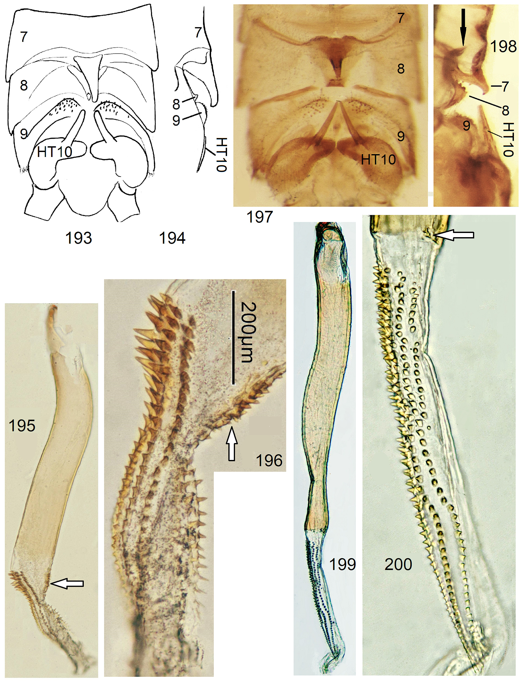
5 T8 with a minute raised hump almost concealed by a long process of T7. HT10 finger-shaped, wing length only ~ 7mm. Penis tube angled near basal third ( Fig. 195 View FIGURES 193–200 ), tip with ventrodistal spine patch (arrow in Fig. 196 View FIGURES 193–200 ) Endophallus on ventral side with a short crest composed of some erect spines ( Figs. 193–196 View FIGURES 193–200 )...................................... 35 N. pusilla n. sp.
5‘ T8 with large raised hump, HT10 a slender straight spike. Penis long, straight, ventrodistal spine patch rudimentary. Endophallus dorsally with long rows of serially arranged spines, ventrally completely bare ( Figs. 197–200 View FIGURES 193–200 )..... 36 N. multiserrata n. sp.
6 Process of T7 short, sometimes button-like, little higher than the process on T8. Dorsal spine band on endophallus much longer than ventral band ( N. dubia -complex)..................................................................... 7
6' Process of T7 pyramidal, about triangular in dorsal and lateral views, projecting beyond front of T8 and located higher than the forward-inclined hump on T8. Remainder variable........................................................... 9
7 HT10 a straight spike with apical button. A large terminal patch of strong spines dorsally on penis tube ( Figs. 318–319 View FIGURES 313–322 )........................................................................................ 56 N. proxima n. sp.
7' HT10 curved, no apical button. External spine patch on penis tube far in front of tip, or weak......................... 8
8 Penis tube with a transverse dorsal belt of spines some distance from tip. Strong spines on knee-like base of endophallus and along dorsal and ventral sides are abruptly replaced by microtrichia covering the endophallus distally ( Figs. 313–317 View FIGURES 313–322 )........................................................................................ 55 N. dubia Klapálek View in CoL
8' External spines on penis weak, scale-like, in a dorsal notch of tube. A band of small spines, no microtrichia, starts far from endophallus base but is long ( Figs. 320–322 View FIGURES 313–322 )................................................. 57 N. vicina n. sp.
9 Endophallus about as long as penis, with wide base, not long and tube-like....................................... 10
9‘ Endophallus much longer than penis tube, narrow, resembling a garden pipe or a rope ( N. spio -cluster)................ 11
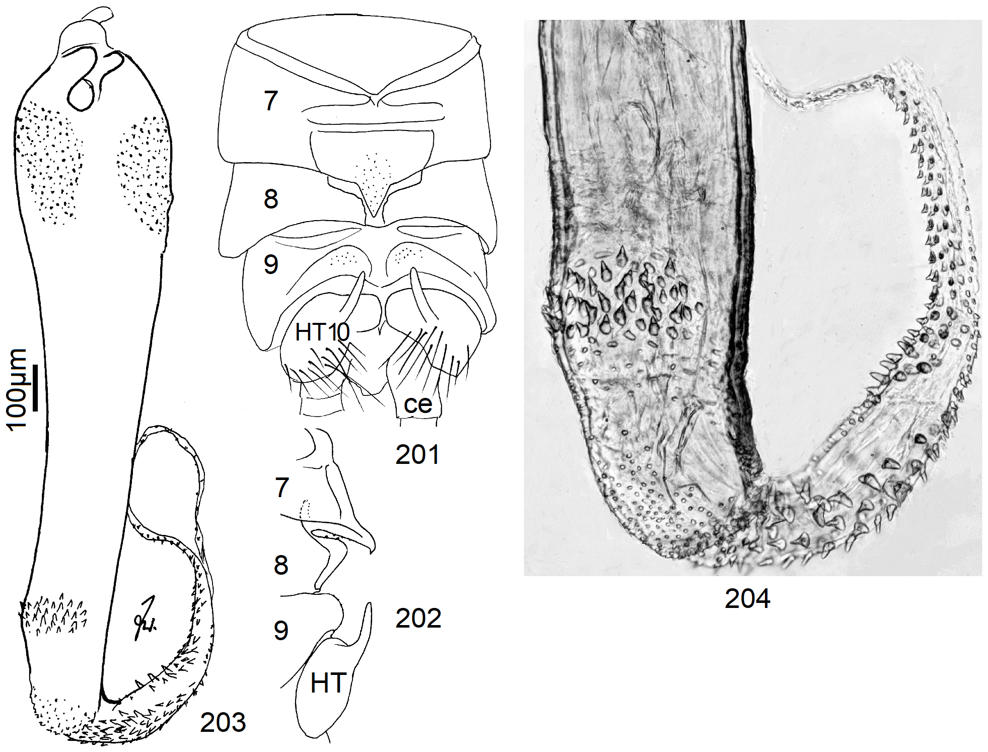
10 HT10 short, finger-like. A dorsodistal patch of spines on penis tube, endophallus short, strongly curved, with wide unordered dorsal and ventral bands of erect cones ( Figs. 201–204 View FIGURES 201–204 ).................................. 37 N. brachyphallus n. sp.
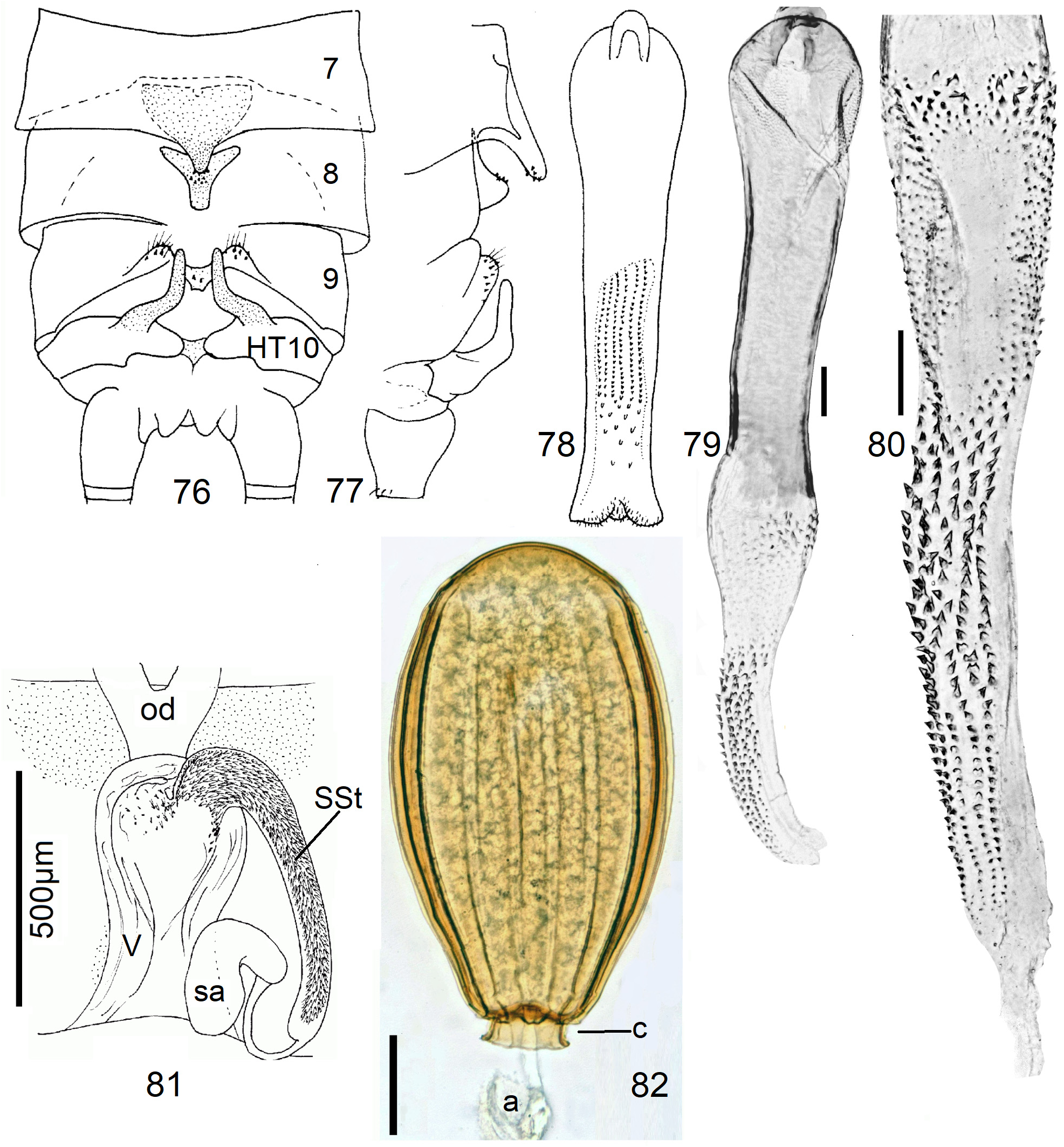
10‘ HT10 longer, angularly bent, tip blunt. Penis waisted, no external spines. Endophallus straight, conical, the wide base with fine spines around a bare center. In the narrow distal part, spines stand in lines ( Figs. 76–82 View FIGURES 76–82 ).......... 1 6 N. duodeviginti n. sp.
11 Strong external spines on penis tube form distinct patches or bands............................................. 12
11‘ No macroscopic spines on outside of penis, dorsal side may bear microscopic asperities............................ 13
12 External spines on penis form dorsal, lateral, and ventral patches. Spines on endophallus distally in three orderly rows ( Figs. 296–300 View FIGURES 294–304 ).......................................................................... 52 N. orthonema n. sp.
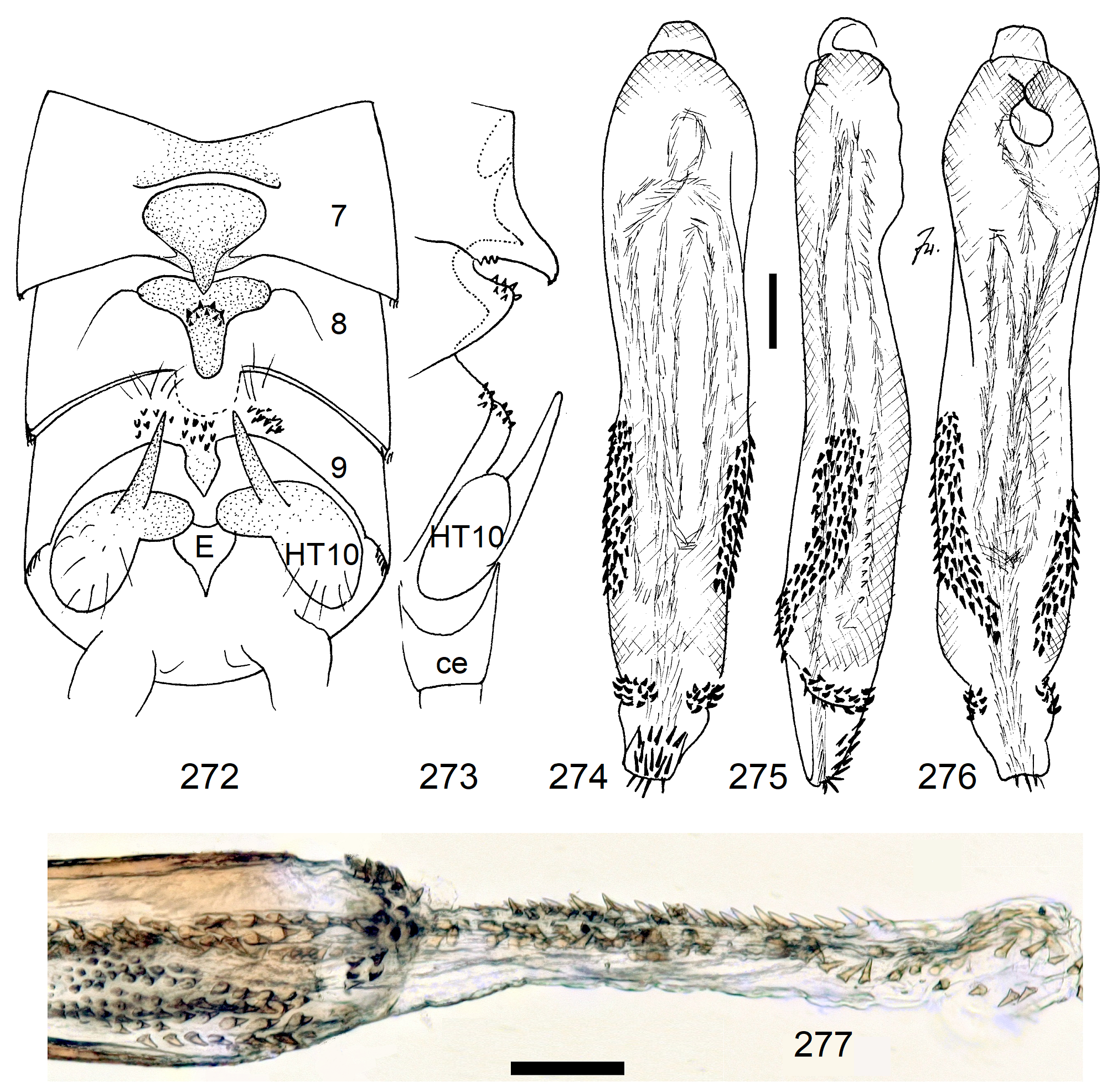
12‘ External spines on penis form winding lateral spine bands. Needle-like spines of endophallus are in no order ( Figs. 272–277 View FIGURES 272–277 ).................................................................................. 49 N. tansanica n. sp.
13 HT10 with truncate or spatulate tip...................................................................... 14
13' HT10 with unmodified tip............................................................................. 16
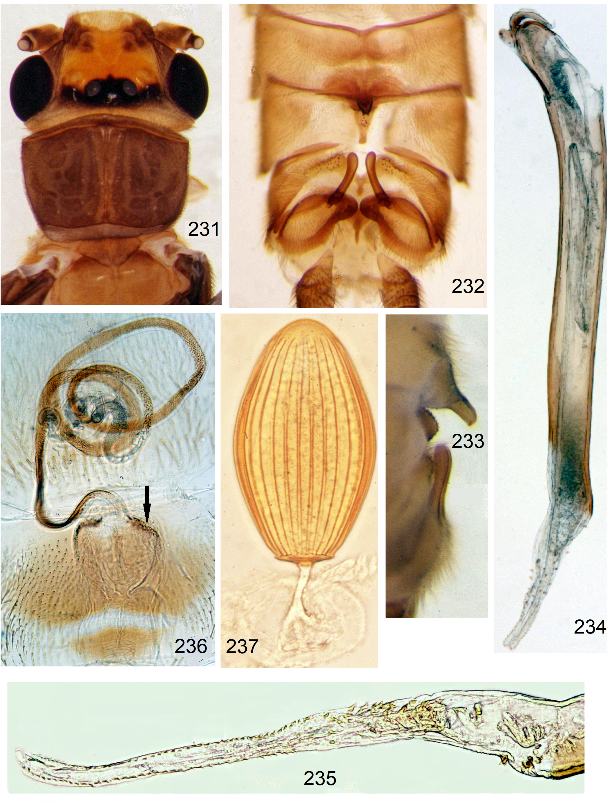
14 Penis is a firm smooth tube almost 9* longer than wide ( Figs. 231–235 View FIGURES 231–237 )..................... 44 N. nigricauda Klapálek
14' Penis tube soft, approximately 5* longer than wide, distal third dorsally with microscopic denticles................... 15
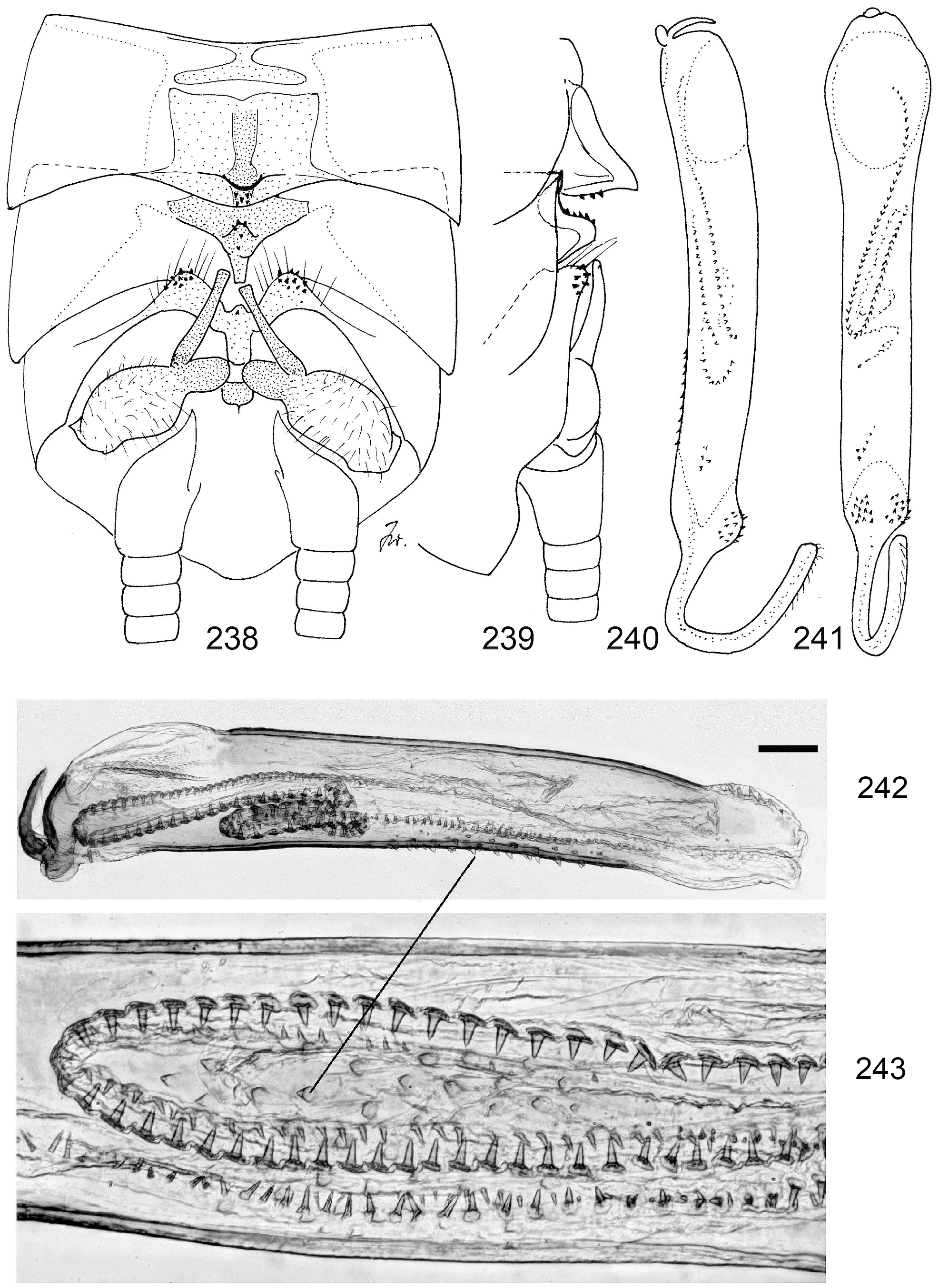
15 Sclerite on T7 with angular front corners, HT10-process straight, apex truncate ( Figs. 238–243 View FIGURES 238–243 ). Wings light................................................................................................ 45 N. spio (Newman View in CoL )
15' Sclerite on T7 with rounded corners, HT10-process short, spatulate ( Figs. 250–254 View FIGURES 250–257 ). Wings fumose....... 46 N. bella n. sp.
16 T8 with a high pointed process almost meeting the process of T7. Penis plump with wide dorso-distal spine patch ( Figs. 351–354 View FIGURES 351–361 )....................................................................... 70 N. conradti (Enderlein) View in CoL
16‘ Penis slender, no dorsodistal spine patch.................................................................. 17
17 Penis apically widened, with few lateral spines............................................................. 18
17 Penis tubular, narrowing towards tip..................................................................... 19
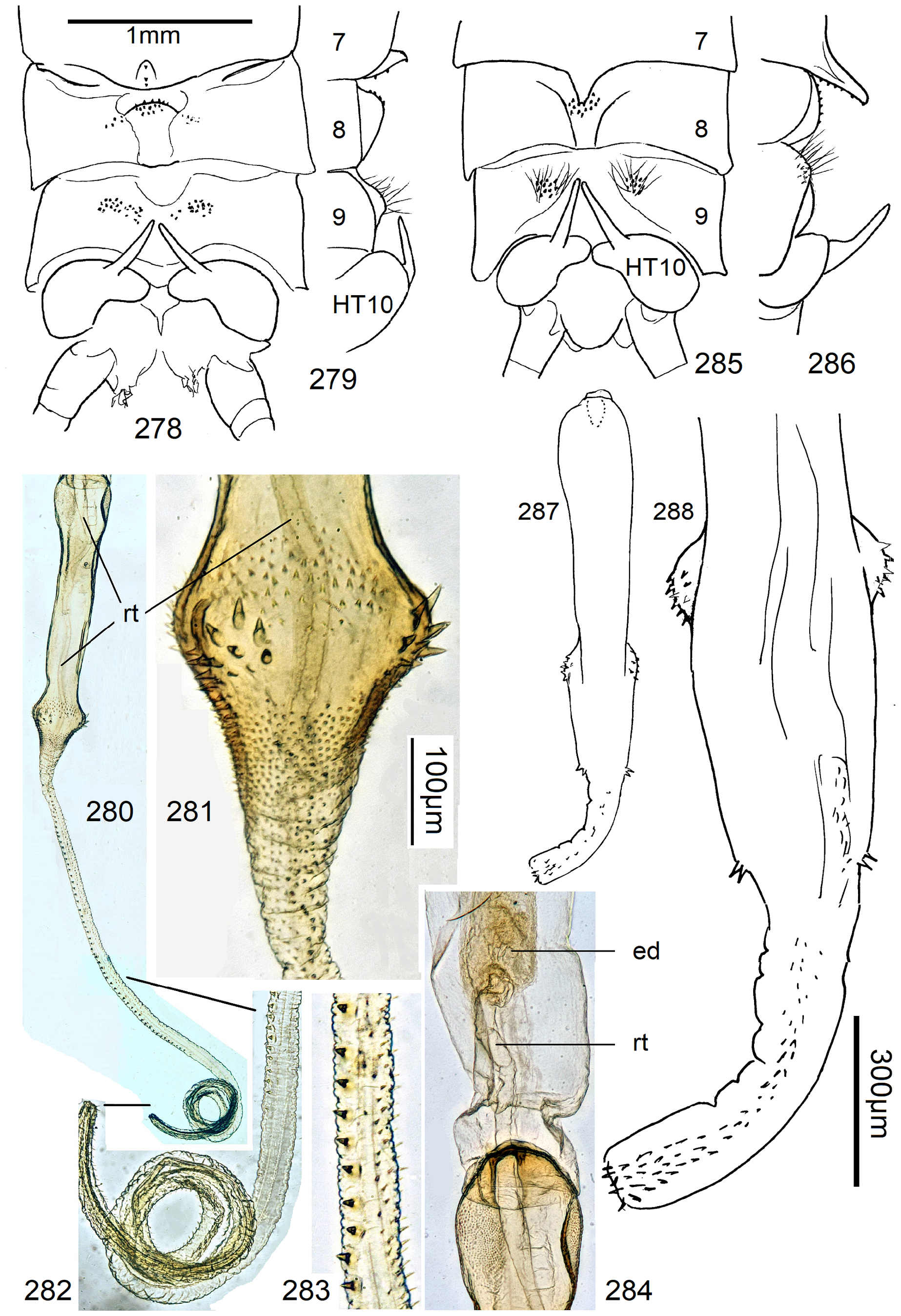
18 Widening of penis short, with few spines ( Figs. 278–284 View FIGURES 278–288 ).................................... 50 N. usambara n. sp.
18' Wide penis apex long, with spines in 4 lateral groups ( Figs. 285–288 View FIGURES 278–288 )........................... 51 N. sambarua n. sp.
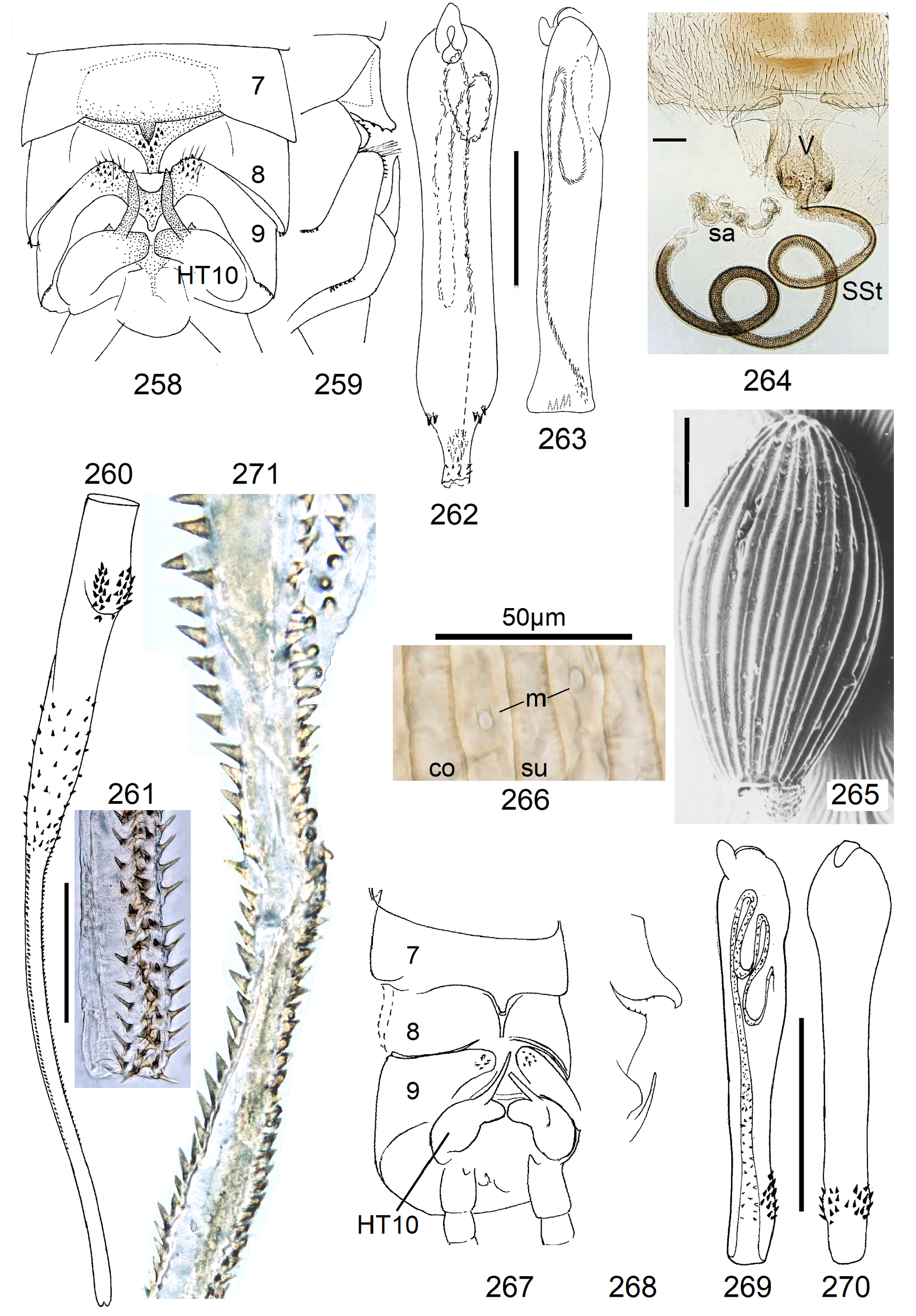
19 Penis tip with few single spines directed caudad. Endophallus a narrow tube with 2 or 3 regular rows of long slender spines ( Figs. 258–261 View FIGURES 258–266 )...................................................................... 47 N. funiculata n. sp.
19' Penis with subterminal spinose swelling, spine tips directed basad. Endophallus base conical, with some delicate spinules, distally a narrow tube with two saw-like rows of teeth (Figs. 267–271)........................... 48 N. biserrata n. sp.
Females
1 SSt straight or bag-shaped, not coiled. ~18 straight egg striae. No marks on S8..................................... 2
1‘ SSt coiled, inside with dense coat of scales, number of egg striae variable. S8 often patterned......................... 3
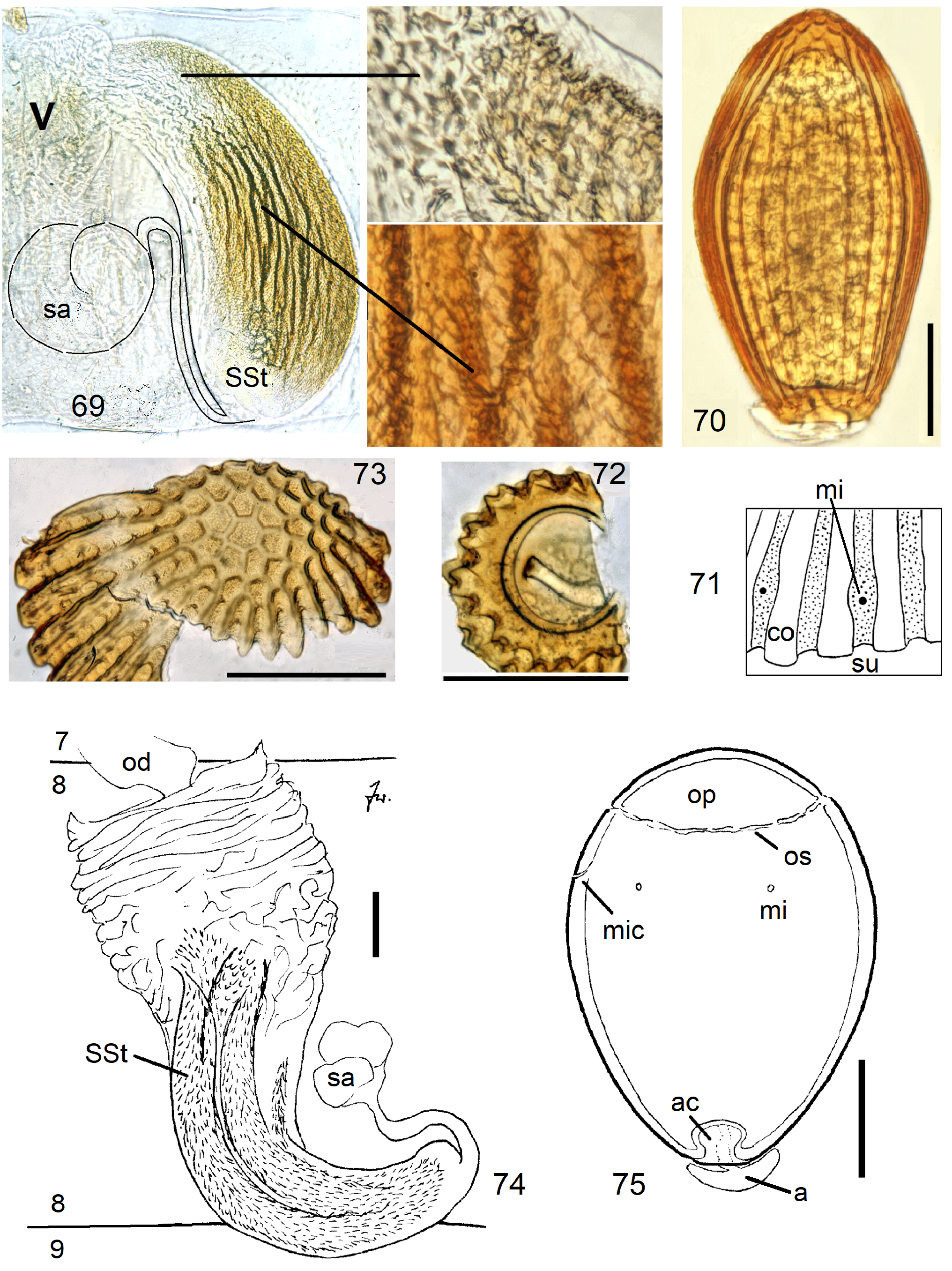
2 SSt finger-shaped, narrow. Egg slender, collar a small crown on the flat anchor pole, operculum convex, punctate ( Figs. 81– View FIGURES 76–82 82).............................................................................. 16 N. duodeviginti n. sp.
2 SSt bag-shaped. Egg ovoid with straight striae, anchor pole not flat, conical operculum with cells ( Figs. 69–73 View FIGURES 69–75 )............................................................................................... 14 N. pickeri n. sp.
3 Two narrow brown marks in a longitudial row on S8. Base and middle of SSt much wider than the terminal fourth. Egg with ~20 striae ( N. dubia -complex)........................................................................... 4
3‘ Terminal section of SSt not narrowing, S8 and egg different, variable............................................ 6
4 Egg with roughly 12 striae, no collar, anchor cavity funnel-shaped, operculum high, conical, with very fine unordered punctation ( Figs. 330–331 View FIGURES 323–331 )......................................................................... 57 N. vicina n. sp.
4‘ Egg with larger number of striae, collar with distinct cells..................................................... 5
5 Costae with ridges or with high flanges which are bent sideways ( Figs. 323–327 View FIGURES 323–331 )................. 55 N. dubia Klapálek View in CoL
5‘ No ridges or flanges on costae ( Figs. 328–329 View FIGURES 323–331 ).............................................. 56 N. proxima n. sp.
6 Anchor cap supported by several fine threads, not by a single stem ( Figs. 301–304 View FIGURES 294–304 )............... 52 N. orthonema n. sp.
6' Anchor cap rests on a single entire stem 6.................................................................. 7
7 Vagina with conical forward curved top containing many scales, the SSt is>10 times longer than the vagina and attaches to the conical top in front.................................................................................... 8
7‘ Vagina approximately calyx-shaped, top not conical, SSt attached dorsally, length of SSt variable...................... 9
8 Egg ovoid, with very narrow sulci and flat smooth costae ( Figs. 289–291 View FIGURES 289–293 )....................... 50 N. usambara n. sp.
8‘ Egg subspherical, not striate ( Figs. 292–293 View FIGURES 289–293 ).............................................. 51 N. sambarua n. sp.
9 Front of S8 with 2 narrowly separated sclerotised plates. SSt many times longer than vagina......................... 10
9‘ S8 different, SSt much shorter, variable, at most 5 times longer than vagina...................................... 11
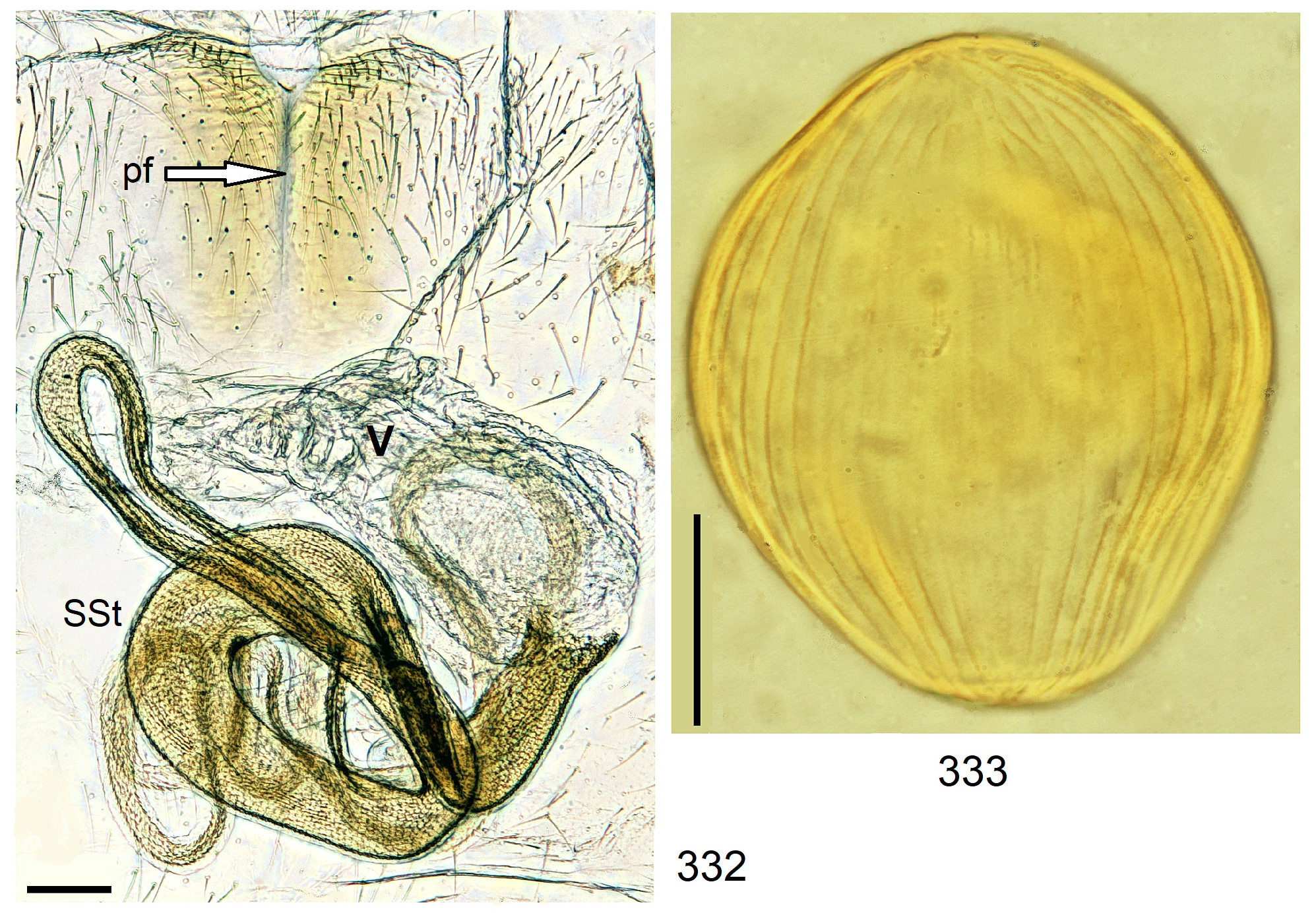
10 Basal part of SSt wide, distal part a very narrow tube ( Figs. 332–333 View FIGURES 332–333 )............................. 58 N. gordius n. sp.
10‘ Entire SSt narrow and tubular, an irregular tangle about as long as the abdomen ( Figs. 334–339 View FIGURES 334–339 )...... 59 N. spaghetti n. sp.
11 SSt stout and short, siccle-shaped, little longer than vagina................................................... 12
11‘ SSt slender, even if short. Egg ovoid, with straight striae, operculum variable..................................... 13
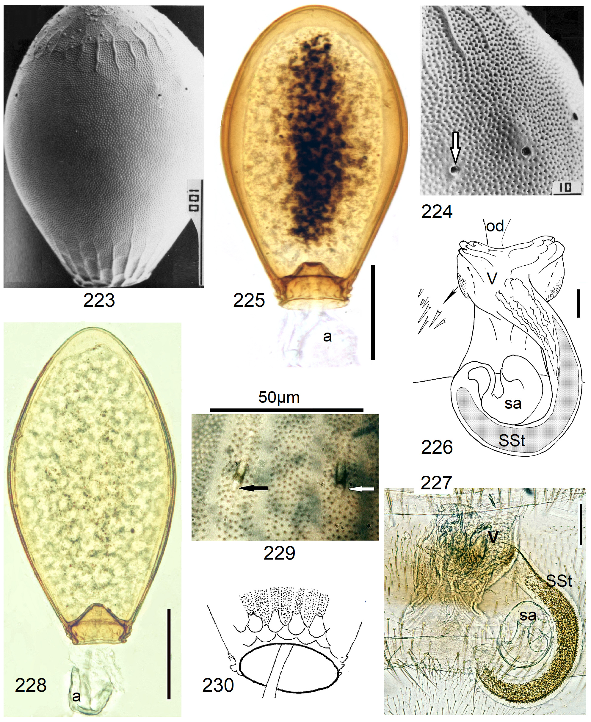
12 Egg subspherical, without striae but near the poles with some short raised wrinkles among coarse irregular punctation ( Figs. 223–226 View FIGURES 223–226 )............................................................................ 42 N. bipolaris n. sp.
12‘ Egg ovoid, slender, the straight striae weakly expressed (Figs. 227–230).......................... 43 N. schuelei n. sp.
13 Groups of spines on both sides of the attachment of SSt to the vagina........................................... 14
13‘ No spines in the vagina laterally from insertion of SSt....................................................... 17
14 About 1/3 of the very long and wide SSt is on the concave side bare, distal 2/3 completely coated by triangular pointed scales. Egg with about 11 straight striae, collar wide and short ( Figs. 244–249 View FIGURES 244–249 )........................... 45 N. spio Newman View in CoL
14‘ Only a short basal section of SSt modified and with scales only on convex side, most of SSt with complete scale cover. More egg striae, or none at all............................................................................... 15
15 On S8 an anchor-shaped pale mark separates a short and narrow transverse arched mark from the large anterior mark. SSt long, tubular. Egg costae and sulci of similar width, both smooth, neither punctate ( Figs. 264–266 View FIGURES 258–266 )........ 47 N. funiculata n. sp.
15‘ On S8 a white arrowhead separates a large anterior brown mark from a short and transverse caudal mark. Egg sulci punctate. .................................................................................................. 16
16 SSt short, forming only ~2 rings. Egg with collar and deep anchor cavity, costae and sulci of similar width ( Figs. 255–257 View FIGURES 250–257 )........................................................................................ 46 N. bella n. sp.
16‘ SSt longer, forming 3–4 rings. Egg without collar and anchor cavity, costae wider than sulci ( Figs. 236–237 View FIGURES 231–237 )............................................................................................ 44 N. nigricauda Klapálek
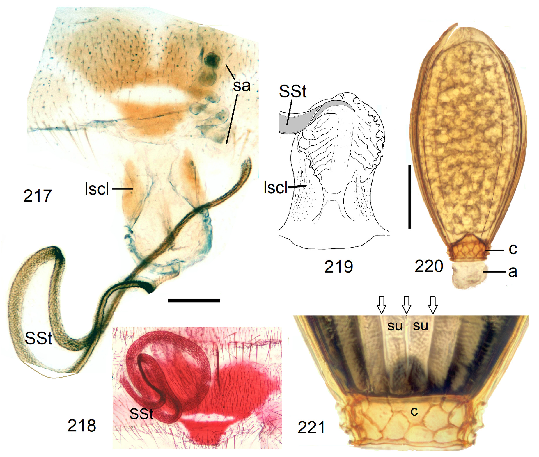
17 Vagina with lateral sclerites, SSt narrow, 2–3 times longer than vagina. Egg costae very narrow, the sulci exceptionally wide. Operculum smooth, no cells or grooves. Caudal brown mark on S9 is a wide truncate triangle narrowly separated from anterior marks on S8 by an oval white window, or a transverse line ( Figs. 217–231 View FIGURES 217–221 View FIGURE 222 View FIGURES 223–226 View FIGURES 231–237 )..................... 40 N. sassandrae n. sp.
17‘ Vagina without lateral sclerites, egg costae and sulci of similar width, or sulci narrrower than costae. S8, SSt and operculum different, variable.................................................................................... 18
18 Three similar separate marks on S8 form a triangle. SSt about 11 times longer than S8 ( Fig. 222 View FIGURE 222 )......... 41 N. benti n. sp.
18‘ Anterior marks on S8 connected, forming a trapezoid. No or only a small caudal mark on S8. SSt variable, from barely twice as long as vagina to several times longer, rarely as long as N. benti . An incompletely resolved difficult complex. Presently only a single new species is formally distinguished from the common and widespread N. burgeoni ......................... 19
19 Anterior half of S8 with large trapezoidal mark, caudal half unpigmented. Egg typically with finely punctate and completely smooth operculum ( Figs. 213–214 View FIGURES 213–214 ) but eggs with delicate ridges surrounding shallow depressions also occur............................................................................................... 38 N. burgeoni Navás View in CoL
19‘ S8 caudallly with a small brown mark. Operculum not smooth but variable, from delicate ridges separating low cells to deep grooves (Figs. 215–216).................................................................... 39 N. beta n. sp.


No known copyright restrictions apply. See Agosti, D., Egloff, W., 2009. Taxonomic information exchange and copyright: the Plazi approach. BMC Research Notes 2009, 2:53 for further explanation.
|
Kingdom |
|
|
Phylum |
|
|
Class |
|
|
Order |
|
|
Family |
|
|
Genus |