
Micrencaustes ( Mimencaustes ) acridentata, Li & Ren, 2006
|
publication ID |
https://doi.org/10.11646/zootaxa.1333.1.4 |
|
persistent identifier |
https://treatment.plazi.org/id/F61D878A-2B1A-FE06-EA28-F55DFDB542DD |
|
treatment provided by |
Felipe |
|
scientific name |
Micrencaustes ( Mimencaustes ) acridentata |
| status |
sp. nov. |
Micrencaustes ( Mimencaustes) acridentata new species ( Figs. 2–14 View FIGURES 2–14 )
Type material
Holotype male, CHINA: Yunnan Province, Xishuang Banna Dai nationality Autonomous Prefecture , N 22°5’ E 101°3’, 16 April 1958, F.L. Li leg. GoogleMaps Paratypes: CHINA: Guangxi Province, Longlin County, N 24°35’ E 104°52’, one female, 10 October 1993, Z.H. Jiang leg. GoogleMaps ; CHINA: Yunnan Province, Jinping County, N 22°56’ E 103°16’, one female, 14 April 1956, K. R. Huang leg. GoogleMaps
Diagnosis
This new species is allied to Micrencaustes ( Mimencaustes) taiwana Araki from Taiwan Province of China. The major differences are listed in Table 1.
Etymology
acri: Latin = sharp, acute; dentate: Latin = spine, tooth. This name refers to the sharp spine at apex of middle tibia.
Description
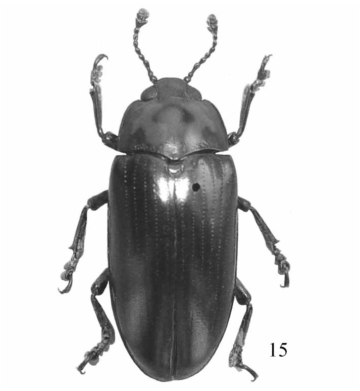
Body moderately elongate, length: 14.0– 15.5mm, width: 5.5–6.0mm (bl/bw = 2.50–2.58; average = 2.54, Fig. 15 View FIGURES 15 ); widest at base of elytra, general color dark, slightly
shining. Pronotum ( Fig. 3 View FIGURES 2–14 ) with two longitudinal, curved orange marks, each bearing a short branch in the middle.
Head ( Fig. 2 View FIGURES 2–14 ) dorsal distance between eyes = 1.87× eye width; ocular striae reaching 0.5 distance to anterior angle of eye; vertex puncture size = 0.5 to 0.75× eye facet diameter, separated by 2 to 3 puncture diameter; epistome puncture size = 1.0× eye facet diameter, separated by 0.5 to 1.0 puncture diameter. Antenna ( Fig. 6 View FIGURES 2–14 ) reaching basal 0.2 of pronotum; antennonere III as long as next 2 antennomeres combined; antennomere IX asymmetrical, almost triangular; antennomeres X to XI symmetrical; antennomere XI subcircular, narrower than antennomere X. Maxillary palp terminal segment ( Fig. 7 View FIGURES 2–14 ) triangular, sides rounded, lateral angle acute, medial angle 90° or more, length = 0.35× width. Labial palp terminal segment ( Fig. 8 View FIGURES 2–14 ) triangular, extended medially, sides rounded, angles nearly 90°, width = 0.86× length; Labial palp width = 0.34× maxillary palp width. Mentum with plate triangular, sides concave; ridge medial extension short and bluntly pointed.
Pronotum ( Fig. 3 View FIGURES 2–14 ) widest at base (pl/pw = 0.71–0.76; average = 0.74); lateral edge slightly curved, strongly margined; anterior edge straight, margined only behind eyes; basal edge weakly sinuate, lacking margin. Pronotum finely punctate, punctures evenly scattered, separated by 3.0 to 5.0× punctures diameter, size = 0.3× eye facet diameter, or less; often with a basal group of coarse puncture, size usually = 1.5× eye facet diameter. Pronotal angle pore present, small; anterior angle pore ventral to marginal line and posterior of angle; posterior angle pore in marginal line; anterior angles projecting.
Scutellum broadly pentagonal, length = 1.87× width.
Elytra (el/ew = 1.68–1.80; average = 1.73), widest near base. Each elytron with 8 complete striae; strial punctures at base of striae VI, VII, and VIII = 2.0× pronotal disc punctures; punctures of striae II, III and IV = 5.0× pronotal disc punctures, gradually becoming finer posteriorly; intervals finely punctured.
Prosternum ( Fig. 4 View FIGURES 2–14 ) triangular between the coxae, produced into a blunt point at the anterior margin, and distinctly depressed in the middle, and emarginated behind; coxal lines straight, length= 0.5× sternal length, lines converging anteriorly and surpassing coxae.
Mesoventrite ( Fig. 5 View FIGURES 2–14 ) broad, with a median quadrate depression; distance between mesocoxae = 1.28× coxal diameter; mesocoxal lines short; sternum with fine and sparse punctures.
Metaventrite broad, distance between meta and mesocoxa = 2.5× mesocoxal diameter; sternum with medial punctures coarse, a few fine lateral punctures.
Mesotibia ( Fig. 15 View FIGURES 15 ) with outer edge of apex acutely toothed.
Male genitalia ( Fig. 9 View FIGURES 2–14 ) with median lobe weakly curved; narrowed to a point; median strut length = 1.28× median lobe length; flagellum short, length = 0.85× median lobe length; sclerite at anterior end of flagellum as in Fig.10–11 View FIGURES 2–14 .
Female genitalia ( Fig. 12–13 View FIGURES 2–14 ) with narrow styli at apex of coxite, and styli rounded apically, covered with setae at apex. Female spermatheca as in Fig.14 View FIGURES 2–14 .
Distribution China: Yunnan, Guangxi.

| R |
Departamento de Geologia, Universidad de Chile |
| VI |
Mykotektet, National Veterinary Institute |
No known copyright restrictions apply. See Agosti, D., Egloff, W., 2009. Taxonomic information exchange and copyright: the Plazi approach. BMC Research Notes 2009, 2:53 for further explanation.
|
Kingdom |
|
|
Phylum |
|
|
Class |
|
|
Order |
|
|
Family |
|
|
Genus |