
Mastogloia ovulum Hustedt 1933
|
publication ID |
https://doi.org/10.11646/phytotaxa.126.1.1 |
|
persistent identifier |
https://treatment.plazi.org/id/F9712264-4D38-FFC5-39B1-FF28D633FE35 |
|
treatment provided by |
Felipe |
|
scientific name |
Mastogloia ovulum Hustedt 1933 |
| status |
|
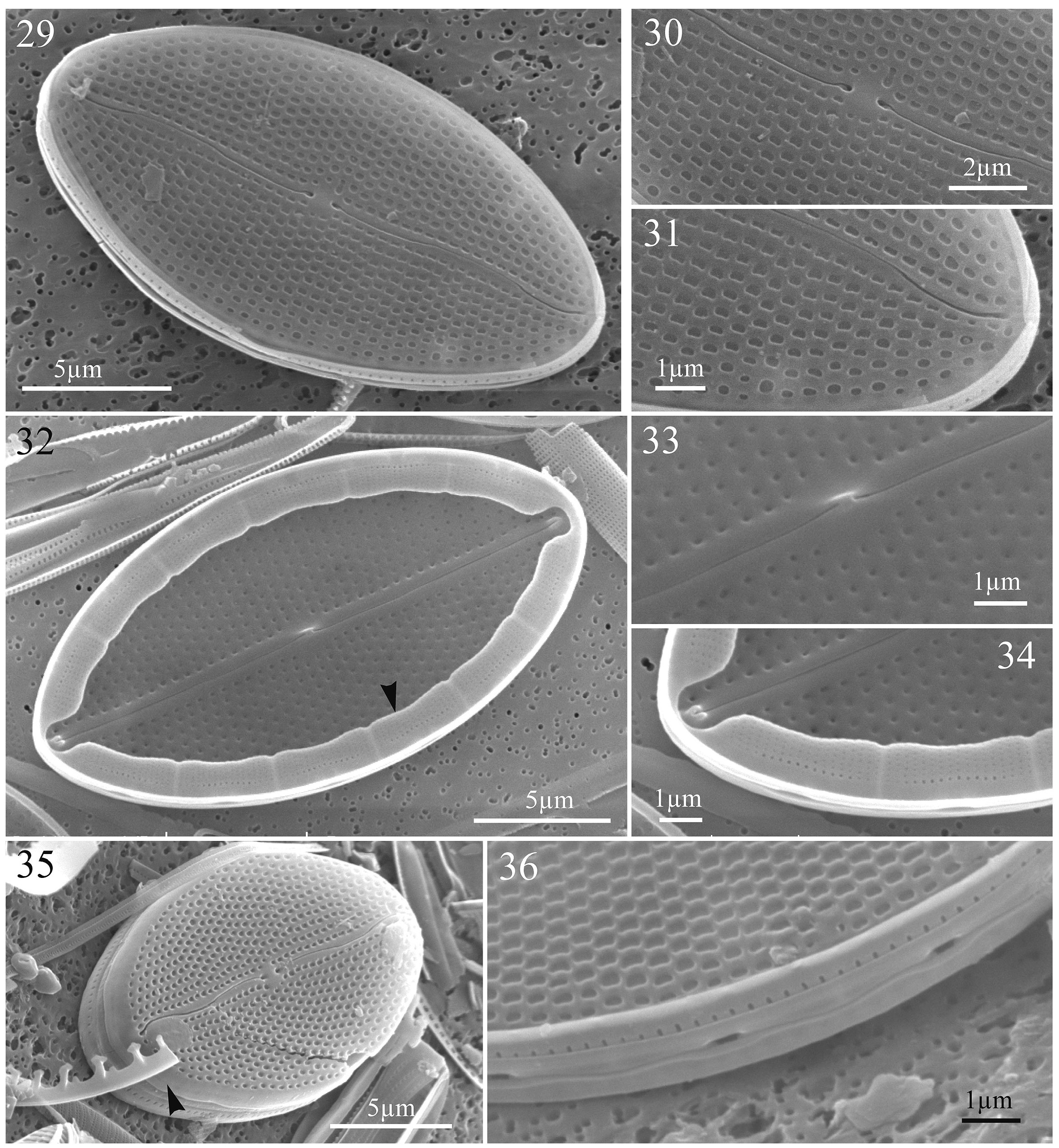
Mastogloia ovulum Hustedt 1933 ( Figs 29–36 View FIGURES 29–36 )
References:— Hustedt 1933, p. 474, fig. 892; non Cholnoky 1968, p. 42, fig. 45; non Montgomery 1978, pl. 141, fig. F (= Mastogloia ovalis ); non Tomàs 1982, figs 18, 19 (= Mastogloia crucicula ); Navarro 1983, p. 121, figs 50, 51; Simonsen 1987, p. 136, pl. 222, figs 1, 2, 6–9, 10?, 11?, non figs 3–5 (= Mastogloia crucicula ); Navarro et al. 1989, p. 352, fig. 70; non Witkowski et al. 2000, p. 255, pl. 75, fig. 14 (= Mastogloia crucicula ); Hein et al. 2008, p. 69, pl. 40, fig. 7.
Material:— Sample from Siladen, Indonesia. SEM stub no. DISVAR-ANS4SP32.
SEM morphology:— The raphe consists externally of two sinuous branches ending centrally in expanded pores and distally in slightly deflected pores toward the same side of the valve ( Figs 29–31, 35 View FIGURES 29–36 ). Internally, the raphe branches run straight and are bordered by ribs, with a rising up central nodule and helictoglossae at poles ( Figs 32–34 View FIGURES 29–36 ). The transapical striae ( 20–26 in 10 µm) are uniseriate and they vary from parallel at centre to radiate at apices, sometimes forming an irregular quincunx pattern ( Figs 29–31, 35, 36 View FIGURES 29–36 ). Externally, the areolae are apically elongated and more or less rectangular in shape, and they are sunken onto the valve surface ( Figs 29–31, 36 View FIGURES 29–36 ), with none observed on the mantle ( Fig. 35 View FIGURES 29–36 , arrowhead). Internally, the areolae open through small rounded foramen ( Figs 32–34 View FIGURES 29–36 ). Partecta slightly bilobed ( Figs 32 View FIGURES 29–36 , arrowhead, 34), equal in shape and size ( 1.1–1.5 µm in width), and occupy the entire length of the partectal ring up to the apex ( Figs 32, 34 View FIGURES 29–36 ). Each partectum is ornamented with small pores in parallel rows ( Fig. 34 View FIGURES 29–36 ) and opens externally through an elongate partectal pore positioned opposite to the concave side of the partectal lobes ( Figs 34, 36 View FIGURES 29–36 ). Length: 17.6–23.4 µm; width: 8.9–10.7 µm ( Table 1).
No known copyright restrictions apply. See Agosti, D., Egloff, W., 2009. Taxonomic information exchange and copyright: the Plazi approach. BMC Research Notes 2009, 2:53 for further explanation.
|
Kingdom |
|
|
Phylum |
|
|
Class |
|
|
Order |
|
|
Family |
|
|
Genus |