
Epanerchodus tujiaphilus, Liu & Golovatch, 2018
|
publication ID |
https://doi.org/10.11646/zootaxa.4459.1.2 |
|
publication LSID |
lsid:zoobank.org:pub:67E956AB-04B1-4EF7-8CC0-E152F95D0563 |
|
DOI |
https://doi.org/10.5281/zenodo.5973766 |
|
persistent identifier |
https://treatment.plazi.org/id/9005EB48-423F-8E5E-48DA-FF740464FEB5 |
|
treatment provided by |
Plazi |
|
scientific name |
Epanerchodus tujiaphilus |
| status |
sp. nov. |
Epanerchodus tujiaphilus View in CoL , new species
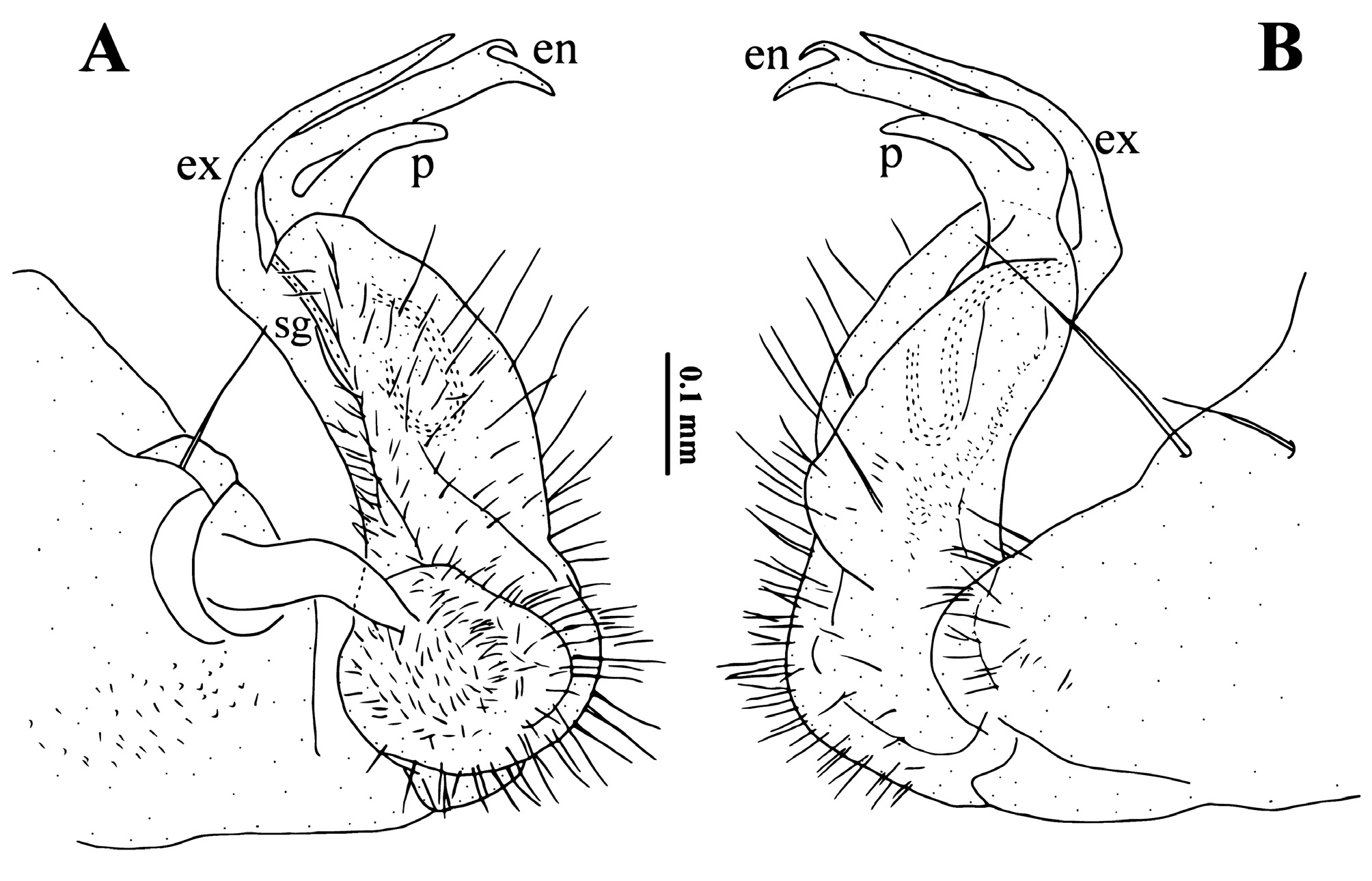
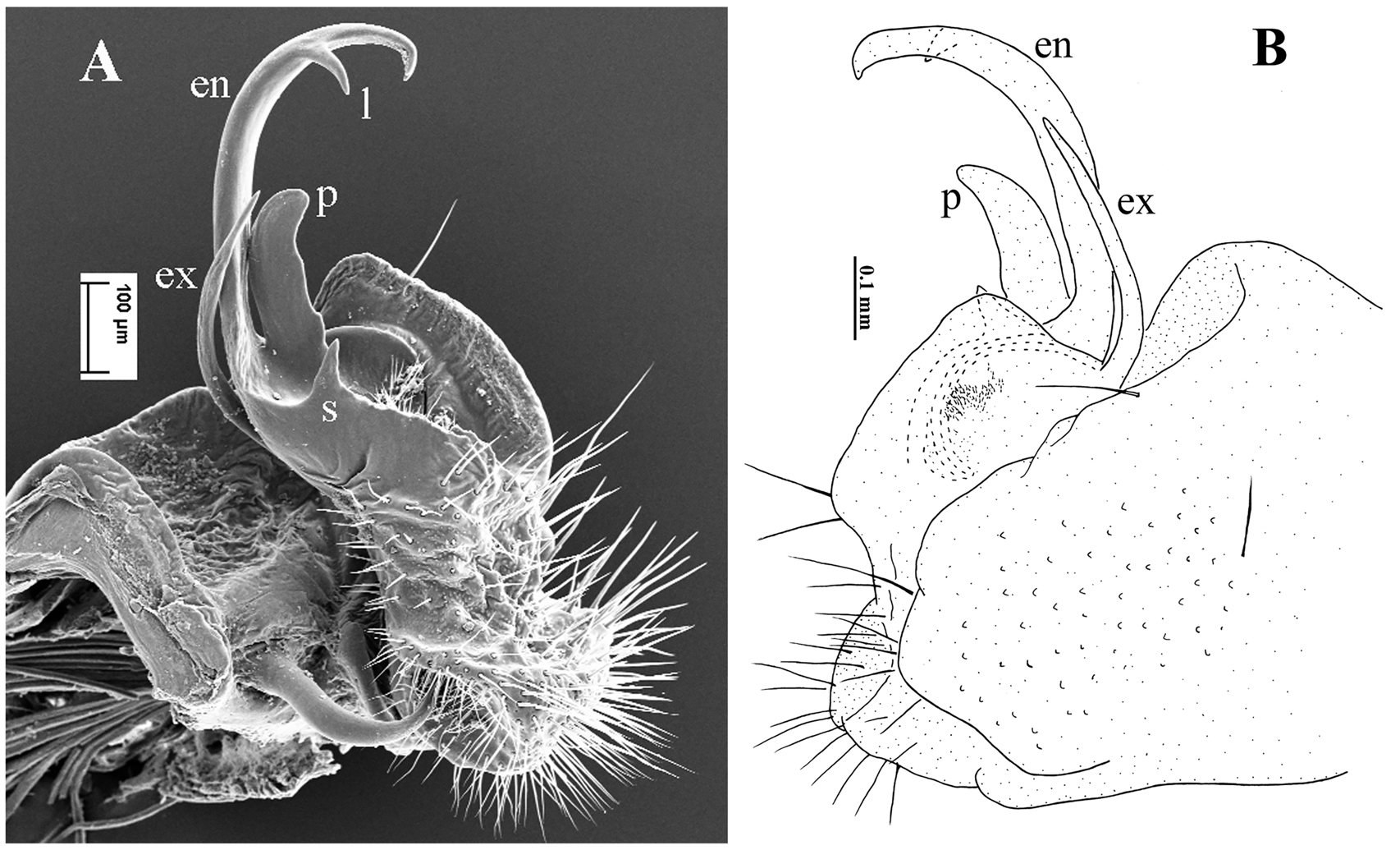
Figs 3–4 View FIGURE 3 View FIGURE 4 .
Material examined: Holotype male ( SCAU eHN2-1) from China, Hunan Province, Longshan County, Huoyan Village, Tujiamei Dong Cave , 2.VII.2014, colls: Mingyi Tian, Weixin Liu, Haomin Yin, Sunbin Huang and Xinhui Wang . Paratypes: 4 males, 9 females ( SCAU eHN2-2), same data as the holotype . 2 males ( SCAU eHN3-1) same data as the holotype, but from different cave: Wulong Dong Cave ; 7 males, 14 females (SCAU eHN4-1), 1 male, 1 female ( ZMUM), same data as the holotype, but from different cave: Panlong Dong Cave ; 3.VII.2014, colls: Mingyi Tian, Weixin Liu, Haomin Yin, Sunbin Huang and Xinhui Wang . 3 males ( SCAU eHN5-1) same data as the holotype, but from different cave: Feng Dong Cave , 30.VI.2015, colls: Mingyi Tian, Weixin Liu, Xinhui Wang and Mingruo Tang.
Diagnosis: Adult males of E. tujiaphiulus n. sp. are distinct from other Epanerchodus species based on the following combination of characters: (1) caudolateral corners of paraterga posterior to collum strongly acute ( Fig. 3A–E View FIGURE 3 ); (2) gonopodal prefemur robust, about 2/3 as long as telopodite; (3) endomere tip unequally bifid; (4) exomere long and spiniform ( Fig. 4 View FIGURE 4 ).
The new species is close to E. varius (from caves in Sichuan, Hubei and Chongqing, China), but is distinguished by (1) the strongly reduced gonopodal femorite devoid of any outgrowths ( Fig. 4A View FIGURE 4 ) vs. with a spine in E. varius ( Fig. 25 View FIGURE 25 ); (2) gonopod with a clearly longer exomere and a broader endomere ( Fig. 4 View FIGURE 4 ) vs. a much shorter exomere and a slenderer endomere in E. varius ( Fig. 25 View FIGURE 25 ).
Description: Based on type specimens. Lengths of body ca 18–22 mm (males) or 20–28 mm (females), widths of pro- and metazonae 1.5–1.8 and 2.8–3.2 mm (males) or 1.6–2.0 and 3.0– 3.2 mm (females). Coloration: in alcohol nearly pallid to light yellowish. Mouthparts light grey-brown, gonopodal telopodites yellowish. Body: Adults with 20 rings. Width: head << collum <ring 2 <3 <4 <5–13, thereafter body gradually tapering posteriorly towards telson. Head: densely pilose, epicranial suture conspicuous ( Fig. 3C View FIGURE 3 ). Antennae long and slender, reaching past anterior margin of ring 4 when extended posteriorly, slightly clavate ( Figs 3B–C View FIGURE 3 ). Exoskeleton: Collum transversely semi-lunar, with an evident lateral incision on each side ( Fig. 3C View FIGURE 3 ). Paraterga evident ( Fig. 3A–C View FIGURE 3 ), midbody paraterga extend metatergite to ca 1.8x width of prozonite. Paraterga 2–7 clearly upturned dorsally above a faintly convex dorsum, other paraterga flat ( Fig. 3B View FIGURE 3 ). Caudolateral corners of paraterga posterior to collum strongly acute, clearly projecting posteriorly past tergal margin. Anterior margin of metaterga bordered and forming a distinct shoulder ( Figs 3A–E View FIGURE 3 ). Integument shining, translucent, prozonae very delicately alveolate. Limbus regularly denticulate. Constriction between pro- and metazonae broad and smooth ( Figs 3A–D View FIGURE 3 ). Metatergal sculpture faint, with three irregular transverse rows of setigerous polygonal bosses. Sulcus between front and middle rows of setae a little deeper than that between middle and caudal rows. Tergal setae visible, short. Three or four faint setigerous incisions at lateral margins of poreless and pore-bearing rings, respectively. Pore formula normal: 5, 7, 9, 10, 12, 13, 15–19, ozopores evident, dorsal, clearly set off from lateral margin and located between last and penultimate marginal incisions. Epiproct long, tip concave, pre-apical lateral papillae evident ( Figs 3D–E View FIGURE 3 ). Hypoproct subtrapeziform, with two setigerous papillae. Pleurosternal carinae absent. Sterna sparsely setose, cross-shaped impressions shallow ( Fig. 3C View FIGURE 3 ). Legs long and slender, about 2.0–2.2 times as long as body ring height in both sexes, without sphaerotrichomes or sternal cones, prefemora not bulging laterally ( Fig. 3C View FIGURE 3 ). Gonopods: subfalcate ( Figs 3F View FIGURE 3 , 4A–B View FIGURE 4 ). Prefemur densely setose and robust, about 2/3 as long as telopodite. Femorite strikingly short, about 1/5 as long as telopodite. Endomere ( en) ribbon-shaped, tip unequally bifid, longest branch only slightly longer than a spiniform, simple exomere ( ex). A single, prominent, digitiform process ( p) at base of, and about half as long as, endomere. Seminal groove ( sg) starting mesally, recurved laterad at base of ex, then turning laterobasad to run into an accessory seminal chamber.
Note: Based on the long slender antennae and legs, and a depigmented cuticle, the species is most likely a troglobite.
Etymology: Derived from the local tribe Tujia ( ± ẎŔ) that populates the area and the Greek “philos”, meaning “liking”; masculine adjective.

| ZMUM |
Zoological Museum, University of Amoy |
No known copyright restrictions apply. See Agosti, D., Egloff, W., 2009. Taxonomic information exchange and copyright: the Plazi approach. BMC Research Notes 2009, 2:53 for further explanation.
|
Kingdom |
|
|
Phylum |
|
|
Class |
|
|
Order |
|
|
Family |
|
|
Genus |